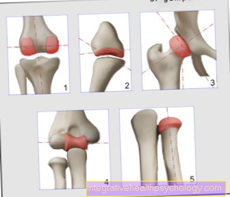
Figure kneecap

- Kneecap -
patella - Femur -
Femur - Shin -
Tibia - Fibula -
Fibula - Inner meniscus -
Meniscus medialis - Outer meniscus -
Lateral meniscus - Kneecap ligament -
Ligamentum patellae - Hamstring muscle -
Rectus femoris muscle - Iliac-tibial tendon -
Iliotibial band - Tibia anterior muscle -
Tibialis anterior muscle
You can find an overview of all Dr-Gumpert images at: medical illustrations
Related images

Illustration
Baker's cyst

Illustration
posterior cruciate ligament

Illustration
Torn knee ligament

Illustration
Inner knee ligament tear

Illustration
Knee joint

Illustration
Back of the knee pain

Illustration
Outside knee pain

Illustration
Inside knee pain

Illustration
Cartilage damage

Illustration
Cruciate ligament

Illustration
meniscus

Illustration
anterior cruciate ligament