Fallot tetralogy
Synonyms in the broadest sense
Congenital cyanotic heart defects with right-left shunt
English: Fallot's tetrad, Fallot's syndrome, Fallot's disease
definition
The Fallot tetralogy is a congenital heart defect. It is one of the most common cyanotic heart defects. Cyanotic means that the heart defect has a negative effect on the oxygen content of the blood. The blood that is pumped from the heart to the organs therefore contains too little oxygen. This is noticeable in the color of the patient's skin. This is pale bluish. The lips in particular appear blue. This type of heart defect has what is known as a right-left shunt. So there is a normally non-existent connection between the right and left hearts.
General
The Fallot tetralogy combines different features of a very specific congenital heart defect. This was first used in 1888 by Etienne-Louis Fallot as a Heart defect described with four different characteristics (Greek: tetra = four):
- Pulmonary stenosis (pulmonary artery obstruction)
- right ventricular hypertrophy (thick layer of muscle in the right ventricle)
- Ventricular septal defect (VSD) (hole in the septum of the heart)
- Crossing the aorta over the VSD
For one thing is the Pulmonary arterywhich the blood from the heart to the lung pumps, stenosed (narrowed / closed). The blood comes from Body circulation relatively low in oxygen in the right heart. First it will be from right atrium into the right ventricle, then pumped into the pulmonary artery from there. If this artery is now narrowed, there is no longer enough blood in the lungs to be reloaded with oxygen.
In some cases, the pulmonary artery can be completely blocked. Then the pulmonary blood flow takes place through a "gait" that persists in the child's development (Ductus arteriosus), which virtually regressively connects the main artery with the pulmonary arteries. Since this normally closes in the first days of life, it is kept open in this case with medication.
Furthermore, the Fallot tetralogy a defect in the heart septum, which usually separates the left from the right heart (medical: Septal defect). The defect lies in the part of the wall that separates the heart chambers from each other (medical: Ventricular septal defect). This is the right-left shunt mentioned above. The blood can now flow from the right heart directly into the left heart. Thus, it bypasses the route through the lungs and is not enriched with oxygen.
The so-called "riding aorta" (Aorta is the main artery in humans) is directly related to the wall defect: Since the septum "has a hole" in the area of the ventricle and the blood can now flow directly from the right ventricle into the left ventricle, this excess amount of blood must also be pumped into the body via the main artery become. Due to this increased pressure, the aorta “rides” over the pulmonary arteries.
The increase in muscle volume (med .: hypertrophy) of the right ventricle is related to the narrowed pulmonary arteries. More force must be used to pump the blood through the smaller diameter of the vessel. Thus the muscle increases in mass; comparable to any other muscle in the body that we train more.
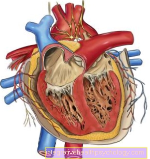
Heart anatomy

- Main artery (aorta)
- ventricle
- Coronary arteries
- Forecourt (atrium)
- Vena cava
- Carotid artery
Summary
The Fallot tetralogy is a congenital heart defect. The existing conditions in the heart create one cyanosis, i.e. an insufficient supply of oxygen to the blood and organs. The children stand out because of their bluish skin. Features include:
- Defect in the heart septum
- Narrowing / occlusion of the pulmonary artery
- Overriding main artery
- Wall thickening of the right ventricle
The disease can express itself with varying degrees of difficulty depending on the severity of the characteristics. The heart defect is usually corrected surgically within the first year of life. Imaging methods such as ultrasound are primarily available for diagnosis. That too EKG may indicate the increased Muscle mass the right ventricle.
Causes of the Fallot tetralogy
The Fallot tetralogy is a congenital heart defect. Embryonic development plays a major role in the development of Fallot's tetralogy. Here the embryo is equipped with all the organ systems that are important for life.
The actual cause of the Fallot tetralogy has not yet been conclusively clarified. However, a genetic disposition is assumed, as some affected children have a chromosomal abnormality, for example children with Down syndrome.
Symptoms
The affected infants have a bluish skin color (main symptom !!), due to the low oxygen content in the blood. However, the extent of the so-called cyanosis depends on the severity of the pulmonary artery narrowing (pulmonary stenosis).With a slight narrowing it can happen that the Fallot tetralogy cannot be identified at all based on the skin discoloration. However, these patients are distinguished by their loud heart murmur. If the lung artery is completely closed, there is a life-threatening undersupply of the entire small organism with oxygen on the 2nd to 4th day of life. Depending on the severity of the oxygen deficiency, so-called watch glass nails and drumstick fingers develop. Watch glass nails are arched fingernails, which got the name because of their shape, which is reminiscent of a watch glass. With drumstick fingers, the end links of the fingers are thickened and appear swollen. Physical development can be quite normal, because as long as the child is still growing in the mother's womb Pulmonary circulation taken over by the mother. The child receives oxygen-enriched blood from the mother because it cannot yet breathe itself.
As a rule, these symptoms appear in just under half of the children in the first 14 days of life. In the majority, however, the symptoms do not appear until the first 3 months of life.
Early symptoms include poor drinking, shortness of breath, and increasing oxygen depletion (cyanosis).
diagnosis
in the EKG typical changes of an increase in muscle mass on the right side of the chamber can be read off. Do an ultrasound of the little one Heart Both the defect in the cardiac septum, the overridden main artery, and the narrowing of the pulmonary arteries can be seen. That too roentgen can provide information. The typical features, such as the enlarged right ventricle, missing pulmonary arteries and thus less vascularization of the lungs can be seen here. If you want to represent the prevailing conditions more precisely, you do one Cardiac catheter. To do this, a thin tube is pushed into the heart through a peripheral vein and contrast agent is injected. This is done with infants under anesthesia.
therapy
The therapy depends on the characteristics of the Fallot tetralogy.
Are the Pulmonary arteriesthat are supposed to transport the blood to the lungs, not only narrowed, but closed, so with the help of medication attempts are made to establish an embryonic connection between aorta (Main artery) and the pulmonary artery (the so-called Ductus Botalli) open. Come here Prostaglandins for use. Prostaglandin E1 is given as an infusion. Is a high grade cyanosis (Lack of oxygen in the blood and the organs to be supplied), the pulmonary artery can be connected to an artery that carries oxygen-rich blood into the body. This is done through a Goretex tube. An expansion of the pulmonary artery with the help of a small balloon is also promising.
The Fallot tetralogy is surgically corrected within the first year of life. The defect of the heart septum is closed so that the main artery (aorta) emerges from the left heart again, as it should normally be. The narrowing of the pulmonary artery is corrected by removing muscle tissue.
prophylaxis

Unfortunately, it is not possible to prevent Fallot's tetralogy. Since the cause has not yet been conclusively clarified, prophylaxis is difficult. Nowadays, however, expectant parents can make use of prenatal diagnostics. Here you can use various methods (from ultrasound to amniotic fluid puncture) to detect congenital deformities, especially heart defects, even before birth (med .: prenatal) determine.
With such a diagnosis it is particularly important that the birth takes place in a specially equipped clinic (e.g .: University hospital or special centers). Because not only are the necessary equipment here, but also appropriately trained and specialized staff on site.
forecast
How this congenital disease progresses primarily depends on the blood flow in the lungs. If this is small, i.e. the defect is large - this means an (almost) completely closed lung artery - so the life expectancy is unfortunately low. Without treatment, every second affected person dies before the age of 20. In the literature, rare cases are also known in which the patients get significantly older.
If the heart defect is corrected surgically, then there is a good life expectancy. The operation itself, of course, comes with its own risks. Bleeding, infections and the like are conceivable here. However, various studies show that the survival rate is little dependent on age. Usually, however, the children are operated on quite early, between 3 and 6 months.
more information
You can get detailed information here information to this topic
- Cardiac arrhythmia
- AV block
- Heart failure
- Anatomy heart
- Atrial flutter
- Heart defect