SI joint osteoarthritis
definition
The ISG, also Sacroiliac joint or called the sacrum joint, is located on both sides of the Basin and establishes a connection between two bones, the iliac bone and the Sacrum, at the SI joint osteoarthritis is it a degenerative wear and tear the articular surface and the Articular cartilagewhich can cause severe pain and restriction of movement in the back and hip area.

root cause
There can be various factors that cause SI joint osteoarthritis. In most cases, SI joint osteoarthritis develops as a result of one Improper loading of Joint between the sacrum and iliac bone. The ISG is at almost everyone Movements of the pelvis involved. Its function is to intercept and reduce the forces generated during movement, and then divide them between the lower and upper halves of the body.
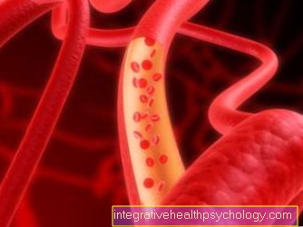
If the load is incorrect, there is a Wear and tear on the articular cartilage. By training an ISGarthrosis The bones that are involved in the structure of the sacroiliac joint can deform and a malalignment of the pelvis can develop. Carrying heavy objects often leads to incorrect stress. Another cause of the development of SI joint osteoarthritis old injuries in the area of the pelvis. These include old pelvic injuries as a result of a serious accidentthat have led to the destruction of the articular cartilage or to malalignment of the pelvis and are also responsible for incorrect loading.
As another cause are Inflammation in the past in the ISG. Chronic inflammation in particular can lead to a remodeling of the joint structures. Also through Obesity The enormous force can put additional strain on the SI joint and articular cartilage. The normal one should also be mentioned age-related degenerative wear of the articular surfacewhich can lead to the development of SI joint osteoarthritis over the years.
Symptoms
Most patients with SI joint osteoarthritis report about strong pain in the deep Back area such as Hip pain and substantial restrictions in the sequence of movements. This pain occurs suddenly when moving up and can radiate into the legs, similar to one Herniated disc of the lumbar spine.
In the early stages of SI joint osteoarthritis, the severe lower back pain initially only under stress, such as standing or walking for a long time. Often times the pain is strongest during the morning hours, improves during the day and decreases in intensity in the evening.
In the process, pain can also occur with smaller movements such as the Bow or even with simple ones Twists of the upper body arise. Also increased sitting can cause severe pain in SI joint osteoarthritis. To escape the pain in these cases a Relieving posture taken to relieve the joint affected by osteoarthritis.
If the SI joint osteoarthritis has existed for a longer period of time, radiate the pain also in the side Pool wall and the Groin area. If the wear and tear of the joint surface and the articular cartilage is very advanced, it often occurs persistent chronic pain one that is only made worse by stress. The malalignment of the pelvis, which is often associated with SI joint arthrosis, can increase over time further complications to lead. To counteract the misalignment and curvature of the pelvis, a curvature of the spine can develop, which further increases the pain and the restriction of movement in the area of the back. Patients with SI joint osteoarthritis are often very limited in their everyday life. Small, everyday movements alone can trigger very severe pain.
Appointment with a back specialist?

I would be happy to advise you!
Who am I?
My name is dr. Nicolas Gumpert. I am a specialist in orthopedics and the founder of .
Various television programs and print media report regularly about my work. On HR television you can see me every 6 weeks live on "Hallo Hessen".
But now enough is indicated ;-)
The spine is difficult to treat. On the one hand it is exposed to high mechanical loads, on the other hand it has great mobility.
The treatment of the spine (e.g. herniated disc, facet syndrome, foramen stenosis, etc.) therefore requires a lot of experience.
I focus on a wide variety of diseases of the spine.
The aim of any treatment is treatment without surgery.
Which therapy achieves the best results in the long term can only be determined after looking at all of the information (Examination, X-ray, ultrasound, MRI, etc.) be assessed.
You can find me in:
- Lumedis - your orthopedic surgeon
Kaiserstrasse 14
60311 Frankfurt am Main
Directly to the online appointment arrangement
Unfortunately, it is currently only possible to make an appointment with private health insurers. I hope for your understanding!
Further information about myself can be found at Dr. Nicolas Gumpert
Osteoarthritis without activation signs
Osteoarthritis is often based on joint inflammation. This is mainly triggered by physical stimuli such as friction and blockages.
Typical signs of inflammation are joint effusions, redness, swelling, pain, overheating and thus restricted joint function. These signs can already be recognized externally in the case of pronounced inflammation. One speaks of an "activated osteoarthritis".
In contrast, there is silent osteoarthritis, in which no activation signs are recognizable. This stage of the disease is often symptom-free. Only clumsiness in movements and stiffness in the beginning of the movement can occur.
localization
Right or left
SI joint osteoarthritis can develop due to the anatomical textures manifest both right and left. Due to malpositions of the Spine or also the hip can be increased Stress on one half of the body arise, whereby the articular cartilage is worn out more on one side than on the other side of the pelvis. In addition to misalignments, especially chronic inflammation be responsible for unilateral SI joint osteoarthritis.
On both sides
SI joint arthrosis that occurs on both sides is less common than one that is limited to just one joint. Misalignments and deformations of the pelvic ring can lead to incorrect posture and incorrect loading of both ISGs. In addition to severe pain, this can be accompanied by an increased Restriction of movement and everyday life. Relief of the bilateral sacroiliac joints is more difficult to achieve than with unilateral impairment.
diagnosis

Since SI joint arthrosis tends to cause unspecific back pain, especially in the early stages, many patients first seek treatment from their family doctor. In most cases, a referral will be made to an orthopedic surgeon.
In order to diagnose SI joint arthrosis, a detailed discussion with the patient is necessary. In this way, specific information about the existing complaints and pain, their location and intensity can be requested. With the help of so-called provocation tests, the doctor can exert stress on the SI joint during certain movements, which can trigger or intensify pain in the case of degenerative or inflammatory changes in the joint. This allows the doctor to gain some information about the exact location of the pain and its spread. In order to assess the bony structures and the joint surfaces, further diagnostics are useful.
In the case of SI joint arthrosis, the worn joint surface and any malpositions and curvatures in the pelvic area can be shown in the X-ray image.
An MRI of the lumbar spine or an MRI of the pelvis is primarily used to classify SI joint arthrosis into different stages. If you only want to assess the SI joint, an MRI of the sacroiliac joint is best. It is also possible to inject locally acting painkillers into the affected joint under X-ray or CT control in order to be able to determine whether the pain subsides under anesthesia.
ICD - 10
If the diagnosis of SI joint osteoarthritis was made by the attending physician, it must be coded according to the ICD - 10 so that it can later be billed correctly by the health insurance company. The ICD - 10 is sold as a Key to naming diagnoses used and based on an international classification system: The diagnosis of SI joint arthrosis is coded with M19.9.
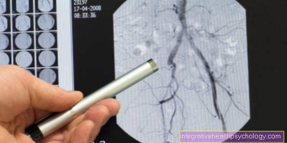
X-ray as a diagnostic tool
In SI joint osteoarthritis, x-rays are a diagnostic method that provides meaningful images, especially in advanced stages of the disease.
With osteoarthritis, there is long-term wear and tear on the joint. In the early stages, x-rays hardly reveal any evidence of osteoarthritis. The changed joint structures can only be recognized late. It is typical that the joint space is reduced and cartilaginous joint surfaces are visibly worn down over time.
The bone also changes steadily over time. Due to the increased friction and wear and tear of the bone near the joint, it rebuilds, which leads to an exacerbation of the disease.
X-rays are also used in therapeutic diagnostics. Painkillers and local anesthetics can be injected into the affected areas under close x-ray control so that the exact location of the pain can be determined. If the pain subsides, the right area has been hit. This also helps when planning further therapies. In rare cases, similar interventions can permanently numb the painful areas. This is a symptomatic but effective therapy.
Vacuum phenomenon in the X-ray or CT image
A vacuum phenomenon is a special feature in the X-ray or CT image that occurs with certain diseases and facilitates diagnosis.
In these radiological procedures, the permeability of individual body tissues to radiation is measured. If, as with SI joint arthrosis, the cartilage in the joint is so worn that it no longer exists and there is a cavity between the bones, this is noticeable in the CT image as a vacuum phenomenon. The gas between the bones is so permeable to the rays that a black hole can be seen instead of the original cartilage tissue. This directly suggests that the cartilage tissue at this point is not intact.
Degree of disability (GdB)
With SI joint osteoarthritis you can severe chronic pain caused. The Pension office can be a so-called for chronic pain Degree of disability pronounce, which varies depending on the disease. The decisive factor for calculating the GdB of SI joint arthrosis is Loss of function in the ISG. Since the function and movement in the SI joint can differ significantly depending on the progress of the SI joint arthrosis, the GdB can vary greatly in patients suffering from SI joint osteoarthritis. With minor functional restrictions, without loss of stability, one can use a GdB of 10 get ranked. At mean loss of function at 20 and only with severe functional limitations and instability can one have a GdB of 30 - 40 to get. However, this can also vary significantly in individual cases.
therapy
Therapy for SI joint osteoarthritis is limited. Those caused by the previous course of the disease Damage to the joint and above all the worn articular cartilage are not reversible. In the foreground is initially one effective relief the symptoms present and especially the persistent pain.
To relieve the pain is one Heat application very useful. Heat can be used in the form of radiation, plasters, fango packs or pillows. Massages can contribute to loosening the surrounding tissue and muscle parts and thereby relieve possible tension.
By special physical therapy the mobility in the SI joint can be improved and possible stress errors and blockages can be compensated. One is also useful Reduction in weight in severely overweight patients. The excess weight exerts more pressure, which leads to a high level of stress on the ISG and can accelerate the process.
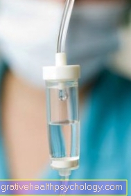
To relieve the pain, so-called non-steroidal anti-inflammatory drugs how Ibuprofen or Diclofenac be applied. Another possible therapy option is infiltration, i.e. the injection of locally effective narcotics. The anesthetics are either injected directly into the joint space of the SI joint or into the ligaments surrounding the joint in order to switch off the pain receptors located there.
Exercises for SI joint osteoarthritis
To the painful course To alleviate SI joint osteoarthritis, the support of a physiotherapist makes sense. Physical activity improves mobility within the SI joint and can counteract tension and possible blockages. Through certain physiotherapy exercises, the joint can be relieved and above all a possible Misalignment compensated become.
One possible exercise for loosening and stretching the ISG is performed as follows: The patient stands with one leg on a stool. The hand on the side of the leg that is on the stool is placed on the Beck shovel placed. With your free hand you should secure yourself on the back of a chair or a table. Then the free leg is swung gently and loosely. After a few repetitions, the side is switched.
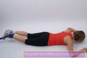
Another exercise is done in Supine position. The legs are lifted and bent so that the lower and upper thighs are at a 90 ° angle to each other. Then the patient makes a movement that the Cycle equals.
To the lower back and especially that Mobilize ISG and to loosen it, you can also put your legs up in the supine position and bend them slightly and place your hands under the sacrum. Then move the pelvis slightly back and forth and move to the side.
There are other simple exercises that should be instructed beforehand by a trained physiotherapist to ensure safe and correct execution.
Surgery for an SI joint osteoarthritis
Surgical treatment of SI joint osteoarthritis should always be viewed as the last option when using more conservative measures There is insufficient symptom relief or there is instability in the joint. In one operation, a stiffening, a so-called arthrodesis. This is a permanent shutdown of the sacroiliac joint.
The surgeon creates suitable access to the SIJ through the skin and muscle layers. after the Joint capsule opened became the damaged articular cartilage Completely away. So that the bony structures that make up the SI joint can be connected to one another, screws or pieces of the body's own bone that were previously removed from the patient's iliac crest are used.
Surgery on this scale harbors some Complications. Next Bleeding, Injuries to the nerves and Muscles can also chronic pain arise and infections are triggered.
For several years there has been a new method that is preferred to stiffen the SIJ (Diana method). With the help of a small incision of a few centimeters in the skin, three titanium rods are installed in the joint via a guide wire, which ensure sufficient stability and grow into the existing bone.
This method is lower risk, has a shorter operating time and a good chance of success. After a stiffening of the ISG you should save for the time being and only partially load the joint until the damaged joint is stable and the nails or titanium rods used are firmly anchored.
Surgical stiffening of the joint (arthrodesis)
Surgical stiffening of the joint should only be considered if conservative therapy is unsuccessful and the osteoarthritis continues to progress painfully.
Nowadays, the procedure can be performed in a minimally invasive manner. The sacrum and iliac bone are screwed together with long screws. The movement of the joint is canceled, which has little effect on the SI joint. The pain is treated long-term and sustained.
prophylaxis

To one SI joint osteoarthritis to prevent it makes sense on one healthy and balanced diet to ensure that there is no excess weight, which is harmful to the bones and joints.
Please also read: Proper nutrition for osteoarthritis
By Smoke the wear and tear of the articular cartilage is promoted. Also is physical activity and adequate exercise good for all joints in the body. This relieves tension and promotes mobilization and blood circulation in the joints.
Specific exercises for the sacroiliac joint can improve the tension, pain and misalignment caused by SI joint arthrosis. Sports that are still recommended are swim or Cycle, as they are especially gentle on the joints. Furthermore, one should pay attention to early symptoms and consult a doctor so that treatment and change of the physical load can take place early in order to counteract a severe and extremely painful course of SI joint arthrosis.


.jpg)