Perthes disease
Synonyms
Legg-Calvé-Perthes disease, ideopathic childhood necrosis of the femoral head
definition
Perthes disease is a circulatory disorder of the child's femoral head of unknown cause.
Age
3-12 years, mainly 5-7. age
Gender distribution
Boys / girls 2: 1 - 4: 1, approx. 15% - 50% on both sides (depending on the source)
Occur
Occurrence approx. 1: 1000 - 1: 5000
root cause
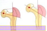
From an anatomical point of view, the blood flow to the femoral head is critical. Most of the blood flow occurs from the Femoral neck, an individually created artery also radiates into the femoral head (see picture on the right).
The cause becomes one Reduced supply of blood vessels accepted. The size of the Circulatory disorder is crucial for the course of the disease Perthes disease and for the regeneration of the femoral head.
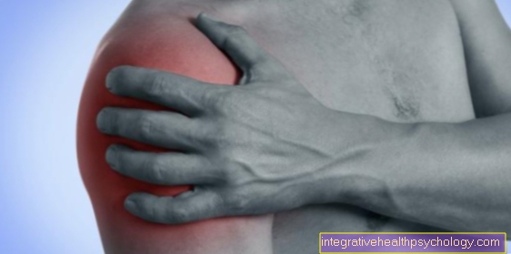
Symptoms
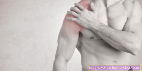
The clinical signs of Perthes disease are initially often only unspecific. In most cases, the child's limp is often the first to be noticed. In the further course, 75% of all children with Perthes disease experience pain in the affected area hip 25% reported pain far from the hip joint knee and Thigh on. The symptoms often change due to the stress and irritation of the joint.
Appointment with a hip expert?

I would be happy to advise you!
Who am I?
My name is dr. Nicolas Gumpert. I am a specialist in orthopedics and the founder of .
Various television programs and print media report regularly about my work. On HR television you can see me every 6 weeks live on "Hallo Hessen".
But now enough is indicated ;-)
The hip joint is one of the joints that are exposed to the greatest stress.
The treatment of the hip (e.g. hip arthrosis, hip impingement, etc.) therefore requires a lot of experience.
I treat all hip diseases with a focus on conservative methods.
The aim of any treatment is treatment without surgery.
Which therapy achieves the best results in the long term can only be determined after looking at all of the information (Examination, X-ray, ultrasound, MRI, etc.) be assessed.
You can find me in:
- Lumedis - your orthopedic surgeon
Kaiserstrasse 14
60311 Frankfurt am Main
Directly to the online appointment arrangement
Unfortunately, it is currently only possible to make an appointment with private health insurers. I hope for your understanding!
Further information about myself can be found at Dr. Nicolas Gumpert
Pain in Perthes disease
Pain is one of the non-specific symptoms of existing Perthes disease.
On the one hand, the dismantling of the joint head causes limp, which gives the first indication of a musculoskeletal disorder or a neurological problem.
The pain often starts with the knee, which can lead to a misdiagnosis. Therefore, Perthes disease should also be considered in children who are free from hip pain, but who have a limp.
If the joint head is gradually destroyed, there will also be more pain in the affected hip. These occur in particular when moving or exercising. A confusingly similar pain picture is shown in the so-called Runny nose (Coxitis fugas). This is also a childhood disease of the hip joint.
A differentiation can optionally be made with the help of an X-ray image.
diagnosis
In the early stages of Perthes disease, the clinical examination can often be normal. As the disease progresses, the hip joint is increasingly restricted in movement. Splaying and rotation in particular are increasingly restricted. As can be seen above, the various stages can be clearly distinguished from one another in the X-ray image. Only in the early phase can the diagnosis of Perthes' disease be made reliably with MRI (magnetic resonance imaging). When comparing the sides, the change in the left femoral head (right side of the picture) is noticeable.
X-ray in Perthes disease
X-rays play an important role in the diagnosis of suspected Perthes disease or in monitoring the progress of a known disease. Only in early detection is the representation by means of an X-ray image inferior to an MRT image of the hip. X-rays can diagnose and classify the disease.
Each stage of the disease of Perthes' disease is shown differently in the X-ray image and can be recognized by experienced radiologists or (pediatric) orthopedists.
In the first stage, the growth plate widens, which is difficult to see on X-rays and can therefore be more easily visualized by an MRI.
In the next stage of condensation, the bone tissue becomes thicker due to the pathological destruction of the substance.
In the picture this shows up as a brightening, as the denser bone structures absorb more X-rays. The destroyed bone is now partially broken down in the fragmentation stage.
The X-ray shows a decomposed head of the thigh bone and the image darkens in the area of the joint due to the decrease in bone structures. In the repair stage, which usually merges seamlessly into the healing stage, the bone is formed again.
The x-ray shows the reconstruction of the joint head and the normalization of the anatomical conditions. If there are deformities during healing because the joint was too stressed in this weak phase, this can also be shown with the help of an X-ray. This pathological change typically shows up as a mushroom-shaped joint head.
Read more on this topic: X-ray examination of the child
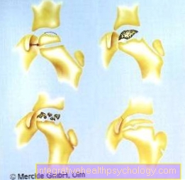
Classification
The classification of the Perthes disease takes place after the four degrees after Cattarall.
The different Catterall stages are classified according to the degree of femoral head involvement. in the Stage I. only a small superficial part of the femoral head is affected.
in the Stage II of Perthes disease are in contrast to Stage I. larger parts of the femoral head have poor circulation. The characteristics can be seen on the left edge of the picture. The Catterall classification must not be interchanged with the Perthes stages. A prognosis of the course of the disease is difficult at any point in time and depends on many individual factors. A maximum of 50% of the femoral head is affected.
in the Stage III of Perthes disease the entire femoral head is affected by the circulatory disorder. Overall, the prognosis is less favorable than the first two Cattarall stages. Unfortunately, at this stage, too, it is not possible to make any predictions about the further course of the disease. A maximum of 75% of the femoral head is affected.
in the Stage IV of Perthes disease if the femoral head is completely destroyed, there is a risk that the remaining femoral head will slip off the femoral neck. An anatomical structure only takes place if the disease occurs at a very young age. The entire femoral head is affected.
Another important classification is that Classification according to Herring. It is of decisive importance for the long-term prognosis. Here the femoral head is divided into three pillars. The outer / lateral pillar is of decisive importance.
Group A: lateral pillar is not affected
Group B: > 50% of the height of the lateral pillar is affected
Group C: <50% of the height of the lateral pillar is preserved, thus the worst long-term prognosis
Stages of Perthes disease
1. Initial stage: This stage is often very difficult to see on the X-ray. Often the growth plate initially only widened. Magnetic resonance imaging (MRI) can provide more information at this stage.
2. Codensation stage: The breakdown of the basic structure of the bone structure results in radiological examination (in the X-ray a compression of the bone structure). This stage is reached approximately 2 to 6 months after the onset of the disease, depending on the severity.
3. Fragmentation stage: After the termination stage, the fragmentation stage follows. Its maximum expression is reached after about 12 months. This stage marks a breakdown of the bone structure, hence fragmentation. Especially at this stage, the femoral head is less resilient.
4. Repair stage: During the repair stage, the femoral head is rebuilt through the sprouting of new vessels. This stage is reached after 2 to 3 years. This allows bone cells to settle again and form basic bone substance. This leads to the reconstruction of the femoral head.
5. Healing stage: The healing stage is the final result of the bony remodeling processes. If the healing takes place in a deformity, that is, in a non-anatomical end rounding of the femoral head, this remains lifelong. This means that there is a great risk of hip osteoarthritis developing. The healing takes place after 3 - 5 years.
therapy
The aim of therapy must be to prevent deformation of the femoral head during the phase of reduced resilience. If deformation has already occurred, the aim must be to restore joint congruence.
Therapy for Perthes disease must always be designed individually and therefore no general therapy recommendation can be made.
If there are no risk factors, a low Catterall stage and a young age, it is sometimes possible to monitor the progress with full stress on the hip joint. Since Perthes disease has a disease course of several years and thus the critical phase lasts for months, consistent therapy of the affected children is often difficult.
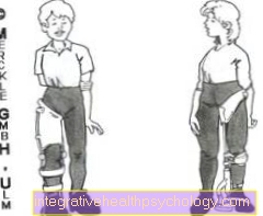
If an unfavorable course is indicated (risk signs according to Cattarall or increasing restricted movement), the hip joint must be relieved by so-called orthoses (see picture on the left) during the critical phase.
If there is a risk of rebuilding the femoral head in a deformed shape, there are various forms of therapy. The aim of all procedures is the so-called containment, the roofing of the femoral head, the decisive stimulation for an anatomical reconstruction, is to be improved. Only two established procedures are mentioned here.
- Correction of the femoral neck straightening with the aim of better centering the femoral head in the acetabulum (intertrochanteric varus osteotomy; IVO).
- Pivoting the roof of the acetabulum using a Salter pelvic osteotomy.
Read more on the topic: Therapy for Perthes disease
forecast
The prognosis for Perthes disease is primarily good. Of course there is no danger to life. Nevertheless, the healing of the disease in an unfavorable deformity of the femoral head can lead to an early hip arthrosis. Basically, however, one can say that diseases before the age of 10 have a more favorable prognosis with regard to deformity, since the body has a higher regeneration potential at a younger age. On the right one can see an unfavorable healing result with typical cylindrical or mushroom-shaped deformation of the femoral head. The original femoral neck is indented, the angle between the shaft and the indicated femoral neck is statically unfavorably too steep. Healing of Perthes' disease in this deformity leads with a very high probability to an early hip arthrosis.
The following are to be assessed as unfavorable factors:
- male gender
- Age of onset> 6 years
- lateral / external calcification in the X-ray image
- pronounced restriction of movement
- Catterall Stadium 4
- Herring Group C
course
The disease of Perthes disease typically has four or five stages.
The course is decisively characterized by the extent of the circulatory disorder.
The individual phases of Perthes' disease are shown below.
There are individual differences in the course that not all stages have to occur in the form and manifestation described in Perthes' disease.
Intertrochanteric varus osteotomy
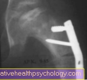
The erection correction of the femoral neck can be recognized. The result was fixed with a plate and screws
Pelvic correction according to Salter
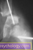
On this x-ray you can see the Salter pelvic osteotomy.
The pelvis is swiveled over the femoral head.
Note
IMPORTANT: The therapy of Perthes' disease belongs in the hands of a pediatric orthopedist! All information is only intended to provide informative assistance.
A Perthes disease can lead to the insertion of a hip prosthesis in old age.


.jpg)


.jpg)