MRI of the hand
General information about MRI
Magnetic resonance tomography (MRT) is based on the magnetic properties of tissue, especially tissue water.
To display the MRI images, a very strong magnetic field is required, which is more than 100,000 times stronger than the earth's magnetic field.
This magnetic field is generated by the MR tomograph.
In addition, it requires electromagnetic waves of high frequency (radio wave range), which have special Do the washing up be generated.
These coils also receive the signal given off by the body tissue. Through the additional application of location-dependent magnetic fields, the signals can be assigned to different body regions and the MRT images can be calculated in this way.
For more information on how the MRI works, please also read: Main article MRI
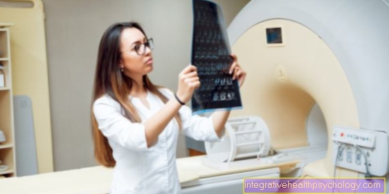
If the safety measures are observed, the MRI represents the hand in contrast to the roentgen a completely harmless examination method represent.
One uses one diagnostically MRI examination on the hand to differentiation between traumatic, degenerative, inflammatory and tumorous diseases of the hand skeleton including the Wrists and surrounding soft tissues or to clarify in general whether there is a pathological finding.
Most often the MRI scan is one X-ray diagnostics upstream.
Disadvantages of the MRI are that high costsassociated with an investigation. It's about four times more expensive than that Computed Tomography and about ten times more expensive than an X-ray examination.
procedure
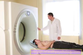
Before a Magnetic resonance imaging can be carried out, it must first be ensured that the patient no ferromagnetic substances in or in itself.
Ferromagnetic materials either trigger a magnetic field themselves or are attracted by an external magnetic field.
This applies to certain implants, Dentures and Pacemaker, which is why such patients are generally excluded from an MRI scan. Due to the ferromagnetic potential, it must therefore be carried out before all items of clothing containing metal, watches, jewelry, etc. are discarded become.
The patient is given a Indwelling cannula into a vein laid over the that Contrast media is fed during the examination.
When performing the handheld MRI, the patient will either in the prone position with your arm stretched out over your head or in the supine positionso that the arm is placed on the side.
For the best possible image quality, the Hand fixed and a receiving coil placed over the hand.
An MRI scan usually takes time between 25 and 30 minutes.
Indications for an MRI of the hand
An MRI examination of the hand or wrist is used for accurate Differentiation of various diseases and injuries. With a precise representation of soft tissue structures (joints, ligaments, muscle tendons), the finest cracks, trauma and signs of wear and tear can be shown. The MRT imaging of the hand is therefore often used after an X-ray diagnosis if no causes of the patient's symptoms could be found in the X-ray images.
Further indications for an MRI of the hand are:
- display of Inflammation
- display of Tumors
- Diagnostics of the rheumatoid arthritis
MRI hand in rheumatism
With the help of the MRI it is possible to suspect a rheumatic disease to confirm or exclude the joints or to assess its course.
In particular, the MRI is used for Early diagnosis used because in roentgen mostly at an early stage no pathological findings finds.
In the MRI you can bony changes recognized earlier and Inflammation in the joint or from Tendons in contrast to X-rays.
For an MRI of the hand in one rheumatic issue is usually for the reasons mentioned above Contrast centerl needed.
Ganglion as an indication for an MRI of the hand
A ganglion (synonym: upper leg) often manifests as a lump in the area of the joint surface. This can occur on both the extensor and flexion side. These are cysts filled with synovial fluid that appear through gaps or tears in the joint capsule. The patient often complains of pain in the wrist, which occurs mainly when there is strong extension or flexion.
Usually, a ganglion can already be visualized in a normal MRI examination. It is an accumulation of fluid in the area of the body surface, which appears white in T2-weighted images. The connection to the actual joint can be made clear by direct injection of contrast medium into the joint capsule (MR arthrography).
Read more on this topic at: Ganglion on the finger, ganglion on the wrist
MRI of the hand for damage to the disc
The Discus of the Wrist (Discus triangularis, TFCC) represents a triangular joint connection between ulna, radius and carpal bones The wrist area can be damaged and lead to pain, especially in the case of torsional loads or injuries in the area of the wrist.
An MRI scan can diagnose the finest injuries in the area of the joint surfaces. In addition, conclusions can be drawn about the development and causes of these injuries.
MRI hand with contrast agent
At a Contrast media it is a substance that strong radiation-absorbing properties possesses, so that an organ or body area through an artificially created density difference better represented can be.
By using contrast media, the Image contrast increased and morbid Blood flow / bleeding of the hand are represented better.
A contrast agent helps to better differentiate between different types of tissue.
For example, without contrast media, muscles and blood vessels are in similar shades of gray shown. Through the Use of contrast media then appear areas with blood circulation on the MRI image of the hand brighter. Pathological processes can thus be detected better and more reliably.
Also Tumors in the area of the hand can be better represented with contrast media, since it often leads to a New formation of blood vessels comes, so that more contrast media then accumulates in tumors.
For example, a usually harmless cyst can be differentiated from a tumor.
It is also noticeable when it is in a tissue less contrast agent accumulatesbecause there is less blood supply to it, for example in scarred tissue.
Before a contrast agent is used, the Kidney values (Creatinine, GFR) to be controlled as the contrast agent again excreted via the kidneys and an accumulation / enrichment of the contrast medium is dangerous for the body.
Control this Laboratory parameters is routinely performed in patients over the age of 50 and patients with known kidney disease.
Before the examination, the patient must three hours sober be, so have not consumed any food or drink.
Especially for Detection of inflammatory diseases a contrast medium should be used, because without a contrast medium, inflammatory processes in joints and tendons cannot be correctly depicted and may be overlooked.
For the Joint displayas it is mostly needed on the hand, is called a contrast medium MultiHance (Gadolinium BOPTA) is used, which is usually well tolerated is.
Gadolinium generally represents that Standard contrast media for MRI examinations represent.
The contrast agent can be used for the examination of joints directly into the joint in question injected, or through a vein (as usual). The joint should then be measured before the MRI scan be moved wellso that the contrast agent can spread out well.
The administration of a contrast agent very rarely results in side effects.
Sometimes it comes to local, usually harmless Side effect reactions at the injection site of venous access.
Because MRI contrast media does not contain iodine are how often X-ray contrast media, it comes side effects are much less common.
It happens extremely rarely allergic reactions by administering an MRI contrast agent. Often it is just one Reddening of the skin.
The frequency of allergic side effects with Impairment of the body's circulation and the breathing occurs at a percentage less than 0.004%.
MRI of the hand without contrast agent
In patients with one Contraindication against the use of a contrast medium, for example by a known renal failure, no contrast agent is used.
Without contrast media can especially bony change be recognized.
Even if there is no contraindication to a contrast agent, an MRI is performed often without contrast media carried out, as it is often sufficient according to the question at hand.
Contrast medium is mainly applied when very similar Body tissue delimited from each other need to be like Muscles and Blood vessels.
For the Detection of inflammation or suspected tumor is an MRI of the hand without contrast agent less suitable.
MRI of the wrist
The term "wrist" is used colloquially for two joints: the joint between the forearm and the carpal bones and the joint between the individual carpal bones. For better presentation of these numerous small articular surfaces with many surrounding ligaments is used for an MRI scan of the wrist.
In the area of the wrist a Rheumatoid arthritis (Rheumatism) manifest. This inflammatory disease can be detected at an early stage with an MRI. A better demarcation is with a Administration of contrast media possible via a vein, as the contrast agent accumulates in the area of inflammation. In addition, various tumors can develop in the area of the bones and the adjacent soft tissue structures. These can also be clearly distinguished from their surroundings with the help of a contrast medium injection.
In recent years, MR arthrography has been used to visualize the various joints. Before the examination, a sterile contrast agent is injected directly into the joint capsule in the area of the wrist under X-ray control. As the joint capsule continues to unfold, the finest cracks in the joint capsule area (e.g. representation of a ganglion), the adjacent cartilage of the joint surfaces (e.g. disc injuries) and neighboring tendons can be distinguished.
Duration of an MRI of the hand
The examination of the hand in the MRI takes time about 30 minutes. However, the duration differs from patient to patient and depending on the question to be examined. With an injection of contrast medium, which is only occasionally required for an MRI examination of the hand, an examination can take a few minutes longer. How often the recording of the images has to be repeated depends on how steadily the hand can be held during the examination. Movements reduce the image quality.
A MR arthrography usually lasts in total up to 90 minutesas the contrast agent must be injected into the joint space approximately 30 to 45 minutes prior to MRI imaging.
costs

Is there a medical indication for performing a MRIs it always is covered by the health insurance.
In general, it can be said that an MRI is actually always taken over if a Transfer a doctor is available for an MRI scan.
Will the MRI only at the request of the patient carried out without the attending physician seeing any indication, the patient must bear the costs yourself.
The doctor should inform them beforehand that the costs must be borne by the patient himself.
For an MRI examination of the hand, the costs are around € 450.
Even if this examination reveals a pathological finding, the costs must be paid ultimately taken over by the patient become.
Do I have to go all the way into the tube?
There are several options for examining the hand. Usually this examination takes place in a closed MRI (colloquially known as a tube). The patient is pushed into the tube with his arm outstretched and fixed. Of the The head and upper body are often still outside the tube.
For a few years there have been specially developed devices that allow an examination of the various joints without the patient having to be pushed into the tube. In the sitting position, the patient extends the affected joint into a magnetic field to be examined.
Do i have to take off my clothes?
For an MRI examination of the hand you have to look usually do not take off. For security reasons, however, if possible all metallic objects put down become. Particular attention should be paid to jewelry and watches in the area of the hand to be examined. These can heat up during an examination and affect the image quality.
In the case of injuries to the hand, a bandage can usually be kept on if it does not contain any metallic structures (e.g. metal splint).




















.jpg)