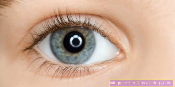
pupil
Synonyms in a broader sense
Eye hole
English: pupil
definition
The pupil forms the black center of the colored iris. Through them the light falls into the inside of the eye, which then goes to the retina (retina) and leads there to the signal transduction, which is responsible for the creation of a visual impression. The pupil is variable in size.
The pupillary reflex is a very important function test in the clinic.

Anatomy & Physiology
The pupil can change its size, this is called pupillomotor activity. It can narrow down to 1.5 mm, which then causes miosis (Greek), its expansion to 8 mm is called mydriasis (Greek) designated.
Two muscles are responsible for pupillomotor function:
- Of the Sphincter pupillae muscle causes the pupil to constrict
- during the Dilator pupillae muscle an extension is obtained.
Both are part of the inner eye muscles. Every muscle needs an innervation from a nerve so that it can be “controlled”. In the case of the muscles for the pupillomotor function, these are nerves of the vegetative or autonomic nervous system. It is roughly divided into two parts, the sympathetic and the parasympathetic. The hallmark of this part of our nervous system is that we cannot or can hardly control it at will. This is also the case with pupil size: the presence or absence of light is primarily responsible for the size. If a lot of light falls on the pupil, the sphincter pupillae muscle is activated. This happens through the parasympathetic nervous system, the pupil becomes narrow. If, on the other hand, it is dark, this leads to an activation of the dilatator pupillae muscle, which is innervated by the sympathetic nervous system, and the pupil becomes dilated.
But besides light as the main initiator for a change in pupil size, other factors also play a role. A classic example is an enlargement of the pupil when you face a person to whom you are inclined. Mydriasis can also occur with excitement and fear. This is due to the fact that in these situations the sympathetic system is activated, which is not only responsible for the eye but also attacks the rest of the body; it is particularly active in situations of increased willingness to act.
A classic example from the time of our ancestors is the "tiger in the bush", the sight of which jumps the sympathetic nervous system and thus optimally prepares people for the upcoming escape. The opposite happens with the parasympathetic nervous system, it is more likely to be active in situations in which one has come to rest.
The pupil size also changes with accommodation (Close-up), here it comes to a miosis, if the opposite look into the distance, the pupil will dilate.
Normally, both pupils are equally spaced (isocoria). If one pupil is significantly wider or narrower than the other, this is called anisocoria. Anisocoria can occur, for example, with increased intracranial pressure (e.g. due to bleeding after a craniocerebral trauma or brain tumors) or in the context of Horner's syndrome, which is classically caused by the triad of miosis (narrow pupil), ptosis (drooping upper eyelid) and enophthalmos (sunken eyeball) is noticeable.
How big are the human pupils?
The size of the human pupil is relatively variable. One of the most important influencing factors is the brightness of the environment. During the day, the pupil has a diameter of about 1.5 millimeters. At night or in the dark, the pupils dilate to a diameter of eight to even 12 millimeters. The circular area of the pupil thus fluctuates between 1.8 square millimeters in brightness and over 50 square millimeters in darkness. With aging, the maximum pupillary opening usually decreases.

- Cornea - Cornea
- Dermis - Sclera
- Iris - iris
- Radiation body - Corpus ciliary
- Choroid - Choroid
- Retina - retina
- Anterior chamber of the eye -
Camera anterior - Chamber angle -
Angulus irodocomealis - Posterior chamber of the eye -
Camera posterior - Eye lens - Lens
- Vitreous - Corpus vitreum
- Yellow spot - Macula lutea
- Blind spot -
Discus nervi optici - Optic nerve (2nd cranial nerve) -
Optic nerve - Main line of sight - Axis opticus
- Axis of the eyeball - Axis bulbi
- Lateral rectus eye muscle -
Lateral rectus muscle - Inner rectus eye muscle -
Medial rectus muscle
You can find an overview of all Dr-Gumpert images at: medical illustrations
Function of the pupil
A narrowing of the pupil causes - similar to a camera - an increase in the depth of field. This is particularly important when imaging close objects. Correspondingly, close accommodation leads to a reflex constriction of the pupil.
In addition, marginal rays are masked out when the pupil is narrow, which reduces blurring due to spherical aberration. The dependence of the pupil size on the brightness ensures that neither too much nor too little light falls on the retina.
The afference runs over the optic nerve (optic nerve, 2nd cranial nerve), which receives the light stimulus, over numerous stations into the Area pretectalis of the midbrain in the brain stem. This is where the efferent path begins, the information becomes a core area in the midbrain, the Nucleus Edinger Westphal both sides, from where the parasympathetic fibers of the oculomotor nerve (3rd cranial nerve) are activated, which at the end lead to a contraction of the Sphincter pupillae muscle lead on both sides and thus to a constriction of the pupil. In the course of the fibers from the eye to the midbrain and back, fibers also partially cross to the opposite side.
Therefore, when one eye is illuminated, not only the pupil of this eye narrows (direct light reaction) but also that of the other eye (consensual light reaction).
With the knowledge of the afferent and the efferent thigh and the fact that normally both pupils constrict when illuminated, conclusions can be drawn about the location of the damage in the case of a disorder of the pupillomotor system:
If the afferent pathway is disturbed (e.g. the optic nerve), there will be neither a direct nor a consensual light reaction when the affected eye is illuminated. When the healthy eye is illuminated, however, both reactions can be triggered. The diseased eye cannot narrow directly, but it can narrow it consensually. This is called amaurotic pupillary rigidity.
If the efferent thigh is disturbed (e.g. the oculomotor nerve), there is no constriction in the affected eye, but a consensual constriction of the pupil on the opposite side, because the perception of the light stimulus (Affinity) is intact so that the healthy opposite side can narrow when exposed to light. If you illuminate the healthy opposite side, the direct light reaction is intact here, but the consensual reaction on the opposite side is not. The affected eye cannot narrow either directly or consensually. This is known as absolute pupillary rigidity
A third disorder of the pupillary reaction is pupillotonia. Here, the pupil of the affected eye is wider when it is bright and narrower when it is dark than that of the healthy eye, whereby the light reaction is slowed down, that is, the enlargement in the dark and narrowing in the case of light is delayed.
The cause is a disturbance of the parasympathetic fibers in the efferent thigh. If the symptoms are also accompanied by a disorder of the muscle reflexes in both of them (In particular, the Achilles tendon reflex cannot be triggered), the disease is also known as Adie syndrome.
The examination of the pupillary reaction is used as standard in almost every clinical examination, it also plays a major role in the diagnosis of coma and brain death.
Pupillary reflex
The adaptation of the pupil to the prevailing light situation takes place via the so-called pupil reflex. A distinction is made between the part that receives the information about the exposure and transmits it to the central nervous system (Affinity) and the part that, after processing this information, leads to the activation of the appropriate muscle (Efference). The illumination of an eye leads to a narrowing of the pupil, this happens via the following structures:
Find out all about the topic here: Pupillary reflex
What is the pupillary distance?
The pupillary distance is the distance between the two pupils. It is divided into the total pupillary distance as well as the right and left pupillary distance. The right and left pupillary distance is the distance between the right or left center of the pupil and the center of the bridge of the nose.
If you add the right and left pupil distance, you get the total pupil distance. The total pupillary distance thus corresponds to the colloquial eye relief.
Usually the pupillary distance is given in millimeters. It is important for fitting glasses and is therefore often included in the glasses passport. When determining the interpupillary distance, it is crucial to look straight ahead. If you look to the right or left, the distance between the center of the pupil and the bridge of the nose changes, of course, and thus also the pupillary distance.
What are the causes of pupils of different sizes?
The size of the pupils is controlled by the autonomic nervous system. It is therefore hardly possible to voluntarily influence it. Normally, both eyes are controlled in exactly the same way, so that both pupils are roughly the same size. This happens depending on the light irradiation. Slight side differences of up to one millimeter are still considered normal.
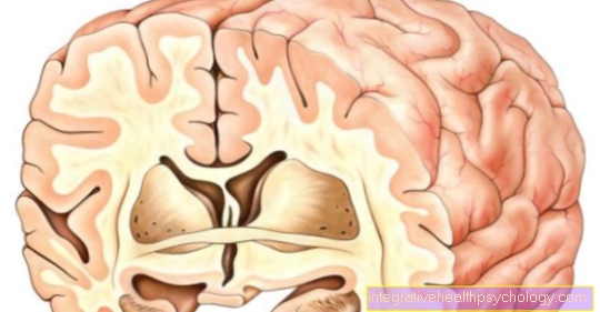
Several parts of the brain are involved in controlling the pupil size. The midbrain is particularly important. From here, signals go to the pupils over several stages. If this area is damaged, pupils of different sizes can be the result. This could be injuries, strokes or cerebral hemorrhage, for example. The signal from the midbrain is sent to the pupils via several interconnections. There may also be disruptions on this route. In so-called Horner's syndrome, part of the autonomic nervous system in the head area fails.
Part of this is usually also involved in pupil control. Since the Horners syndrome often occurs unilaterally, the pupil can be disturbed on one side, resulting in uneven pupils. In addition, the upper eyelid hangs down on the affected side and the eyeball appears sunken. Other causes of pupils of different sizes can be, for example, disorders of the muscles that adjust the pupil size or increased intracranial pressure.
Read more on the topic: Different sized pupils- these are the causes
What can dilated pupils indicate?
When it is dark, the pupils are dilated to allow as much light as possible to enter the eye. The so-called sympathetic nervous system dilates the pupils. It is particularly active in stress reactions and also increases heart rate and blood pressure, for example. In stressful situations, the pupils can be dilated accordingly.
A negative situation does not necessarily have to be responsible for this.
Even with pleasant stimuli, such as looking at a loved one, the pupil also appears to expand. How strong this effect is is, however, controversial in science. Various substances, including prohibited intoxicants, can dilate the pupils. These include, for example, cocaine and methamphetamines. But the ophthalmologist may also give special drops during certain examinations, which dilate the pupils.
Read more on the topic: Dilated pupil
What can constricted pupils indicate?
In bright light, the pupil is narrowed to reduce the amount of light entering the eye. But the pupil can also be narrowed for other reasons. For example, the pupil seems to narrow when looking at pictures that are perceived as disgusting or uncomfortable.
Narrowed pupils could be detected even with very high mental exertion. Whether and how much the pupil narrows or widens due to different situations or stimuli is, however, controversial in science.
Excessive fatigue can be a trigger for constricted pupils.
The pupil can also be narrowed in various diseases. Usually there is damage to the brain areas that are responsible for controlling the pupil. These include meningitis or strokes. In the so-called Horner syndrome, the control of the pupil is also disturbed. The nervous system is no longer able to dilate the pupil of the affected eye, the pupil appears narrowed. Finally, there are a number of substances that can constrict the pupil. These include various pain relievers such as morphine, but also certain eye drops, for example against glaucoma.
Read more on the topic: Horner syndrome
How do the pupils change with drug use?
Many drugs also affect the size of the pupil. The reason for this is that the pupil size is controlled by parts of the nervous system that can react strongly to given drugs. Some drugs can even act directly on the eyes and affect the pupil size there. A basic distinction can be made between substances that dilate the pupils and those that constrict the pupil.
Substances that dilate the pupil are typically stimulants such as cocaine or amphetamines. Both substances work through a similar mechanism. They increase the concentration of the messenger substances noradrenaline and adrenaline in the synapses. This has an activating and euphoric effect on the nervous system. In the eye, however, noradrenaline and adrenaline have a pupil-expanding effect.
Substances that constrict the pupils are typically opioids such as heroin or strong pain relievers. They have a rather dampening effect on the nervous system. Certain parts of the nervous system cause the pupil to narrow when exposed to opioids. Even if the pupil can react to drugs, the pupil size alone cannot clearly determine whether a person is under the influence of drugs.
Further interesting information on this topic can be found at: Which drugs or drugs affect the pupil?
Read more on the topic: The consequences of drugs
What does "isokor" mean for the pupil?
Pupils are called isokor if their diameter is roughly the same on both sides. Slight side differences of up to one millimeter are still referred to as isokor.
Larger differences are no longer isocorative; such a condition is called anisocorus. Since anisocore eyes is an important symptom in a number of diseases, the doctor often pays attention to whether the pupils are isocorative.
Learn more at: Anisocoria
Clinical fact
Medication can be used to intervene in the pupillomotor system. For medical purposes in the context of ophthalmological examinations, for example, drugs are used that lead to mydriasis.
Usually these are administered in the form of eye drops.After it was explained above that the sympathetic nervous system is responsible for a dilatation and the parasympathetic nervous system for a narrowing of the pupil, it is now understandable that one can either give medication to activate the sympathetic nerve in order to achieve mydriasis (Sympathomimetics) or those that inhibit the parasympathetic nervous system (Parasympathetic agents). Usually there is a second application, including atropine and tropicamide.
The induction of miosis can also be medically desirable, for example in the case of an acute glaucoma attack (glaucoma). The intraocular pressure is greatly increased here, the aim is to reduce the pressure as quickly as possible in order to prevent permanent damage to the eye. By constricting the pupil, the aqueous humor in the eye can flow away better, whereupon the intraocular pressure drops. Common miotics are carbachol and aceclidine, both belong to the group of parasympathomimetics, i.e. they activate the parasympathetic nervous system.
Opioids such as Morphine cause miosis. This can be an indication of opioid intoxication in an unconscious patient.
For more information on a pupillary difference, see Different sized pupils.
Twitching pupils - what could be behind this?
There can be many causes for pupils twitching. A pendulum-like tremor of the pupils is also known as nystagmus. It can be congenital or occur as part of a disease such as a visual impairment, brain damage or balance problems. Nystagmus can also occur in healthy people. By jumping back and forth, you can, for example, look closely at objects from a moving train or keep your surroundings in focus when turning around your own axis.
Read more on the topic: Nystagmus
A slight twitching of the pupils is sometimes also described by healthy people when they are very tired and have to focus on one point for a long time. For example, this can occur when working on a screen for a long time or when viewing a lecture. This twitching is probably not dangerous and is related to eye fatigue.