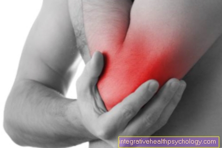
Pain in the little finger
definition
The little finger of each hand consists of three finger bones (phalanges), the basal, middle and distal phalanx. The metacarpophalangeal joint connects this with the metacarpal. The middle and end joints of the fingers lie between the individual finger joints. These joints are surrounded by joint capsules. Tendons and muscles that can be controlled by finger nerves ensure the mobility of the little finger. All of these structures as well as the surrounding soft tissue and the nail area of the little finger can individually or together be the starting point of pain in the little finger. The pain stimuli can have different qualities, for example pressing, throbbing, stabbing, shooting in or electrifying. The pain can be acute or persistent and can vary in severity.
Causes of Little Finger Pain
Possible causes could be:
- Bone cyst
- Injuries
- Inflammation of the nail bed
- arthrosis
- Dupuytren's disease
- Avulsion of the extensor tendon
The causes are now explained in more detail below
Pain in the little finger can originate from bony structures - a break in one of the finger bones, a hairline crack or a bone cyst, as well as a tumor of the bone can be the cause. In the case of a broken bone, the exact start of the pain can usually be linked to a fall or an accident. Cuts and other wounds in the area of the little finger can also be painful. The tendon sheaths of the little finger can be inflamed and therefore painful when moving. Inflammation of the nail bed can also affect the little finger and are often noticeable as throbbing pain and additional signs of inflammation.
Shooting, tingling, or electrifying pain indicate damage or irritation to a nerve or the nervous system in general.
The joints of the little finger can also be affected and cause pain in the case of inflammatory changes, for example rheumatism and gout or signs of wear and tear (osteoarthritis). The so-called Dupuytren's disease leads to flexion contractures, especially on the little finger, due to knotty and hardened tendons on the inside of the hand. If blood vessels or nerves are irritated, it can lead to pain in the little finger. In general, acute overstrain or improper strain on the little finger can also lead to pain.
You may also be interested in this article: Sore Finger Joints - The Most Common Causes
Inflammation of the nail bed
The inflammation of the nail bed or nail wall is also known as paronychia. There is often painful swelling and reddening of the affected nail area. Sometimes pus is already draining. In advanced infections, fever and chills can also occur. If the inflammation spreads to deeper structures such as tendon sheaths, it can lead to so-called phlegmon, a purulent infection of the soft tissues. This must then be treated with antibiotic infusions and, if necessary, surgery. One tries to avoid this urgently. Infection can occur during manicures or when biting your nails, as well as with an ingrown nail, but bacteria can also be introduced into the nail area through cuts and bites. An inflammation of the nail bed often has to be surgically repaired (for details see therapy).
This article might also interest you: Inflammation of the nail bed on the finger
Appointment with a hand specialist?
I would be happy to advise you!
Who am I?
My name is dr. Nicolas Gumpert. I am a specialist in orthopedics and the founder of .
Various television programs and print media report regularly about my work. On HR television you can see me every 6 weeks live on "Hallo Hessen".
But now enough is indicated ;-)
In order to be able to treat successfully in orthopedics, a thorough examination, diagnosis and a medical history are required.
In our very economic world in particular, there is too little time to thoroughly grasp the complex diseases of orthopedics and thus initiate targeted treatment.
I don't want to join the ranks of "quick knife pullers".
The aim of any treatment is treatment without surgery.
Which therapy achieves the best results in the long term can only be determined after looking at all of the information (Examination, X-ray, ultrasound, MRI, etc.) be assessed.
You can find me at:
- Lumedis - orthopedics
Kaiserstrasse 14
60311 Frankfurt am Main
Directly to the online appointment arrangement
Unfortunately, appointments can only be made with private health insurers. I ask for understanding!
Further information about myself can be found at Lumedis - Dr. Nicolas Gumpert
Pain in the little finger after an accident
If pain occurs in the little finger after an accident, a possible bone fracture and an injury to the soft tissues must be clarified. Open wounds are easy to diagnose. If there is also an obvious bone fracture in the depth, it is an open fracture. Otherwise, a closed fracture must be excluded, for example by means of an X-ray. If you fall on the edge of your hand or get trapped in a door, the bones of your little finger can break. Hairline cracks in a bone (fissure) can also cause pressure and strain pain. It is also important to recognize where the fracture is in the bone: If this also affects adjacent joints, the therapeutic procedure can change. Nail fractures or bruises under the nail should also not be overlooked.
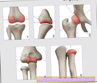
Heberden osteoarthritis
Heberden osteoarthritis is the wear and tear of the finger end joint, for example the little finger (also distal Interphalangeal joint (Called DIP). You feel pain, and you can feel the knotty thickening of the joint on the back of the finger. The joint can also assume a misalignment and deviate to the thumb side. The strength and mobility of the finger can decrease significantly.
Women are more often affected than men; the disease usually occurs during menopause. There is no other cause, the disease is primarily genetic and / or hormonal. Permanent, e.g. Occupational overload should also be considered, as this can loosen the joint capsule, among other things.
Read more about this under: Finger osteoarthritis
Bouchard osteoarthritis
In contrast, Bouchard osteoarthritis affects the median joint of the finger (the proximal interphalangeal joint (PIP)) and is also noticed by pain and nodular thickening at the affected joint. Here, too, there can be swelling, restricted movement, axial deviations and instability. This form of osteoarthritis is rarer than Heberden's osteoarthritis. Overall, however, both forms of osteoarthritis are widespread, especially in older people. Men and women are equally often affected by Bouchard osteoarthritis. Often a patient suffers from this arthritis of several fingers at the same time.
Avulsion of the extensor tendon
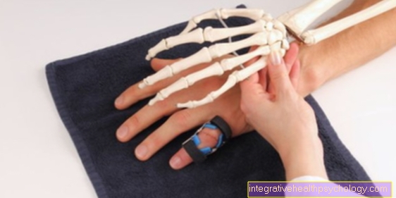
If the little finger is forcibly bent too much, the extensor tendon at the end of the finger can tear off. This can happen in ball sports. A bony tear with an additional fracture of the phalanx is also possible. Therefore, an X-ray of the finger should be taken. Active extension of the phalanx of the little finger is no longer possible after the extensor tendon is ruptured. The resulting permanent flexion is also known as the "hammer finger". You may also notice swelling and tenderness. Splint or surgical treatment are possible options.
You can find more information here: Tearing of the extensor tendon of the finger
Sulcus-Ulnaris Syndrome
The sulcus-ulnaris syndrome or ulnar channel syndrome describes a pain syndrome of the ulnar nerve on the elbow.
The ulnar nerve is one of the main nerves of the arm and, like the median nerve, extends into the hand. Among other things, he is responsible for the sensitivity of the little finger and ring finger. If in the course of this there is bumps, pressure or blows on the nerve, an unpleasant tingling pain occurs along its entire course up to the little finger.
The nerve on the elbow is very superficial and exposed. Its position and the typical pain that is triggered when bumping into this area is also known as the "musician's bones". If the ulnar nerve is repeatedly impacted and stressed in this area, the nerve can be permanently damaged, which can lead to chronic pain in the little finger with weak muscles in the affected hand muscles.
Loge de Guyon syndrome
The Guyon Lodge is an anatomical area on the wrist. Similar to the carpal tunnel, the Guyon box forms a passage over the carpal bone through which nerves and blood vessels can move from the forearm to the hand.
The anatomical tightness of the wrist can lead to compression of the ulnar nerve, which in turn can result in pain in the little finger and ring finger, as well as muscle weakness in the area of the little finger. Cyclists or construction site workers are most commonly affected. The tightness and thus the Loge de Guyon syndrome can also be favored by fractures in the wrist or ulna.
More on this topic: Loge de Guyon syndrome
Therapy of pain in the little finger
In general, short-term pain relievers such as aspirin, diclofenac or ibuprofen can be taken for pain in the little finger. If the little finger is cut, it must be sewn and bandaged, depending on the length and depth, otherwise a plaster is sufficient. If the X-ray shows a fracture that is not displaced or near the joint, it can be immobilized with a plaster cast for several weeks. If there is a displaced or near-joint fracture, the broken bone is fixed in the correct position in an operation using a plate or nail. In Heberden's osteoarthritis, cortisone injections can be made into the affected joint to slow down inflammatory processes. Surgery often has to be considered. In this the joint is stiffened (arthrodesis) and thus no longer causes pain. Only the targeted flexion of the wrist then fails. Targeted, regular movement of the joint can slow down the progression of osteoarthritis. With activated osteoarthritis, cold treatments, otherwise heat applications, can be helpful. The same general therapeutic principles apply to Bouchard osteoarthritis, but surgery is rarely performed. In the case of an inflammation of the nail bed, the finger will be immobilized, elevated, protected and cooled if there are minor findings, and the affected nail area will be disinfected. In addition, an antibiotic is sometimes given. As soon as there is a fever or an abscess is observed, an inflammation of the nail bed must be treated surgically: the area is pierced, thoroughly rinsed and a drainage inserted. In addition, antibiotic therapy is always initiated. Tetanus protection should also be checked, especially in the event of injuries. If the extensor tendon is ruptured, an accompanying fracture or an open injury requires an operation with tendon sutures, otherwise a splint (e.g. Stack’s splint) is put on to keep the finger end joint in extension. This is worn for four to six weeks. Tendonitis can either be treated with sparing and infiltration of cortisone, or if this is unsuccessful, it can be split lengthways in an operation to make more space for the tendon. When diagnosing Dupuytren's disease, the hardened cords can be loosened in an operation if the movement restrictions are extremely stressful in everyday life. Rheumatism drugs can provide relief from joint problems affecting the little finger.
Accompanying symptoms with pain in the little finger
In addition to pain in the little finger, other signs of inflammation such as overheating, impaired functionality, swelling and redness may be noticed. Then a targeted search should be made for the inflamed area, for example a joint or the nail bed. Perhaps individual movements can no longer be carried out, a failed finger extension, for example, indicates a torn extensor tendon, as well as a no longer feasible flexion to a flexor tendon injury. If there is a disease or injury to a nerve, the finger can feel numb. If the blood supply is compromised, the little finger can become cold and pale, for example if the artery is compressed or severed.
additional swelling in the little finger
Swelling of a joint of the little finger or the whole finger is a common symptom in addition to pain. A swelling can indicate an inflammatory or a traumatic event. As a typical symptom of inflammation, joint inflammation such as rheumatism or inflammation of the nail bed can be the cause. Osteoarthritis of the individual joints is also possible. In the case of a bone fracture, additionally injured vessels often lead to a bruise and thus to swelling and blue discoloration. It is important to know the exact location of the swelling in order to rule out or diagnose certain causes.
You may also be interested in this article: Joint swelling on the finger
Pain in the metatarsophalangeal joint of the little finger
In the joint between the phalanx of the small finger and the metacarpus, pain can occur with rheumatic diseases, with wear and tear of this joint (arthrosis) or with an acute gout attack that manifests itself in this joint. In rheumatism, in addition to pain, other signs of inflammation such as redness and overheating are in the foreground. The wear and tear of the joints can be accompanied by the formation of nodules and also lead to swelling. If gout is suspected, the uric acid levels in the blood should be checked, as the accumulation of crystallized uric acid in the joint triggers the acute symptoms.
Pain in the middle joint of the little finger
The Bouchard osteoarthritis mentioned above occurs in the respective middle finger joints, including the middle joint of the little finger. This can cause pain in the middle joint as well as rheumatism or gout disease.
Pain in the femoral joint of the little finger
The terminal joint of the little finger can be affected by what is known as Heberden's arthrosis, which is associated with the formation of knots at the corresponding joint. Here, too, other possible causes are rheumatic inflammation of the joint or a gout attack. Processes that affect the nail, such as nail bed inflammation, can also radiate into the neighboring joint due to the spatial proximity.
Pain in the joint capsule
The joint capsule completely surrounds a joint and thus forms a closed space around the joint itself. Joint effusion can occur within a joint capsule due to increased synovial fluid production. This then leads to pain and restriction of movement of the joint. Joint diseases such as injuries, inflammation and signs of wear and tear also affect the joint capsule directly. In the event of a trauma, the joint capsule can tear, causing stabbing pain and swelling of the joint, sometimes also a bruise.
Duration of pain
The duration of the pain depends on the underlying cause. If it is only a matter of incorrect or overloading, the pain should have disappeared after a few days if you take it easy. In the case of a broken bone, the pain should soon be over after correct treatment, but the duration of treatment is in the range of weeks. Diseases such as gout or rheumatism can be brought under control with medication, but they can also flare up again and again. If nail bed inflammation is treated correctly surgically, this should also quickly lead to relief. After operations, depending on the severity, pain sensations in the little finger can be expected for a few days.
Special features when bending
If pain occurs particularly when bending, the flexor tendons may be affected - possibly caused by irritation due to incorrect or overloading or inflamed tendon sheaths (tendovaginitis). Diseases in the area of the joint can also lead to limited and painful flexion, such as osteoarthritis or rheumatic disease.
Diagnostics for pain in the little finger
The diagnosis should always begin with a detailed questioning of the patient (anamnesis). In such a conversation, questions are asked about the time of the pain, a possible accident, the exact location of the pain and changes in movement, the quality of the pain (pressing, dull, stabbing, electrifying, etc.) and any accompanying symptoms. It is often also important to know more about the patient, such as the job they do or similar symptoms / illnesses in relatives. Next, the affected little finger must be thoroughly examined, paying attention to signs of inflammation, injuries and mobility as well as misalignments. If a bone fracture is suspected, an X-ray must be taken to confirm it. In the case of inflammation or a rheumatic disease, certain blood values can help. In addition, ultrasound and magnetic resonance imaging (MRI) exams can provide clarity.