Stages of femoral head necrosis
General

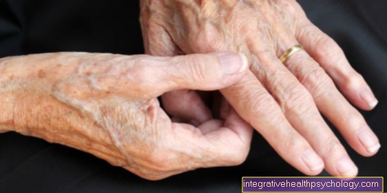
At a Femoral head necrosis the bone tissue of the femoral head dies due to insufficient blood flow (ischemia). Causes for the Circulatory disorder injury to the hip joint, various diseases, Cortisone- and chemotherapy, Irradiation as well Obesity and Alcohol abuse be.
Metabolic disorders, alcoholism or trauma can lead to the development of femoral head necrosis.
The "sinking" of parts of the bone results in a deformation of the femoral head. As a result, it no longer “fits” optimally into the joint socket, causing pain and increasing pain Hip joint wear (Coxarthrosis).
There are two different stages for the course of femoral head necrosis: The ARCO classification divides femoral head necrosis according to the joint changes visible in imaging procedures, while the Ficat / Arlt classification also takes into account the symptoms that occur.
Ficat and Arlet stages
The staging of femoral head necrosis according to Ficat / Arlet includes the findings of the X-ray image also include the patient's symptoms:
Stage 0: The patient has no complaints.
Stage 1: When exercising, pain occurs in the groin area, the hip joint is slightly restricted in movement.
Stage 2: The X-ray image shows the first changes such as the formation of cysts on the femoral head or sclerotherapy (increase in bone tissue below the articular cartilage).
Stage 3: The patient has pain even at rest, the movement of the hip joint is moderately to severely restricted. The x-ray shows that parts of the cartilage have died, leading to deformation of the femoral head.
Stage 4: The femoral head has collapsed and the cartilage shows signs of wear from osteoarthritis.
Appointment with a hip expert?

I would be happy to advise you!
Who am I?
My name is dr. Nicolas Gumpert. I am a specialist in orthopedics and the founder of .
Various television programs and print media report regularly about my work. On HR television you can see me every 6 weeks live on "Hallo Hessen".
But now enough is indicated ;-)
The hip joint is one of the joints that are exposed to the greatest stress.
The treatment of the hip (e.g. hip arthrosis, hip impingement, etc.) therefore requires a lot of experience.
I treat all hip diseases with a focus on conservative methods.
The aim of any treatment is treatment without surgery.
Which therapy achieves the best results in the long term can only be determined after looking at all of the information (Examination, X-ray, ultrasound, MRI, etc.) be assessed.
You can find me in:
- Lumedis - your orthopedic surgeon
Kaiserstrasse 14
60311 Frankfurt am Main
Directly to the online appointment arrangement
Unfortunately, it is currently only possible to make an appointment with private health insurers. I hope for your understanding!
Further information about myself can be found at Dr. Nicolas Gumpert
Stages according to ARCO
The ARCO classification of the Femoral head necrosis was developed in 1992 by the Association Research Circulation Osseous. It assesses the stages of femoral head necrosis according to the changes that can be recognized during imaging diagnostics. The following radiological methods are used:
- roentgen
- MRI (Read more about this topic at: MRI of the hip)
- CT (Computed Tomography)
- Scintigraphy
The patient is injected with weakly radioactive particles that accumulate at the pathological joint changes and are made visible with a special camera. In this way, femoral head necrosis may be detected in the early stages before it becomes visible in the other imaging procedures.
Stage 0 (initial stage): No changes are visible in any of the imaging procedures. Only a microscopic examination of a tissue sample (histology) shows evidence of dying tissue (necrosis).
Stage 1 (reversible early stage): X-ray and CT images are normal, whereas the MRI and / or scintigraphy show evidence of a pathological change in the joint (unspecific change / storage).
Stage 2 (irreversible early stage): X-ray and CT images now also show a change in the joint. In the MRI it is already clearly visible that tissue in the hip joint is dying, although the contour of the femoral head is still preserved.
Stage 3 (transitional stage): A fracture of the femoral head below the articular cartilage can be seen in X-ray and CT images (subchondral fracture). An incipient deformation or flattening of the femoral head may also be visible.
Stage 4 (late stage): All imaging procedures show the onset of osteoarthritis with a narrowing of the joint space and damage to the joint socket.
Therapy based on the stages
Depending on the stage classification according to ARCO, the treating orthopedic surgeon decides which therapy to use Femoral head necrosis it is a possibility:
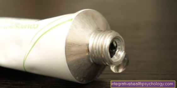
Early stages: In stages 0 and 1, the joint can be relieved by using crutches in conjunction with physiotherapy and anti-inflammatory pain relievers like Ibuprofen or Diclofenac be successful. Medicines with the active ingredient iloprost have a vasodilating effect and stimulate blood circulation and can thus lead to an improvement in the clinical picture. In addition, physical therapy methods such as a Shock wave therapy or magnetic field therapy.
Advanced stages: In stages 1 and 2, core decompression is the standard therapy. In one operation, the femoral head is drilled and so the pressure in the Bone marrow lowered. This can prevent or at least delay the progression of the femoral head necrosis. It may also stimulate repair mechanisms and the formation of new vessels. In modern therapeutic approaches, the body's own stem cells are injected into the bone as part of the drilling, where they are supposed to support the formation of new bone.
- Late stages: In stage 3 and 4 there is often a so-called Corrective osteotomy carried out. This operation is performed on Femoral neck an artificial fracture is created and the femoral head is "repositioned", ie rotated out of the stress zone. As a result, it "fits" better into the joint socket and the damaged bone is relieved. However, this method only works if there is still sufficient healthy bone tissue. If, on the other hand, too large parts of the femoral head are affected by the necrosis, only joint replacement through one can help artificial hip joint (total endoprosthesis, TEP).
Further information on the treatment of femoral head necrosis can be found at: Femoral head necrosis therapy
MRI for femoral head necrosis
In order to be able to make the diagnosis of aseptic, non-traumatic femoral head necrosis and to divide it into a certain stage, the following are usually imaging procedures necessary.
A common division into 4 stages is the ARCO classification (Association Research Circulation Osseous), which is based on an X-ray or MRI examination becomes possible.
- Stage 0 This is the case when changes in the hip joint cannot be detected either in an X-ray examination or in an MRI image, but symptoms are present and the beginnings of the femoral head necrosis can be confirmed by means of a histological evaluation.
- In stage 1 the first changes (in the form of unspecific additional enrichment of contrast media in the femoral head area) in the MRI, but not in the X-ray image. This is due to the fact that the MRT, with its sharper and more detailed display options, allows pathological structural changes to be recognized much earlier than an X-ray image. However, stage 2 can then be detected both in the MRI and in the X-ray, the femoral head contour is preserved in both imaging, although it can already be clearly seen that tissue is dying (necrosis).
- It is different in stage 3in which there is a fracture (Burglary) below the femoral head cartilage.
- Stage 4 is finally characterized by the fact that the femoral head is clearly flattened in the MRI and no longer looks normally round, the joint space is narrowed and the first signs of a Hip osteoarthritis are recognizable.