Talar fracture
introduction

Of the Talus (Talus) is next to that Calcaneus (Heel bone), Navicular bone (Scaphoid bone), Ossa cuneiformia (Cuneiform bones and the Os cuboidem (Cuboid bone) part of the tarsus (Tarsus). The talus forms with its upper side, the Trochlea tali (Joint roller), part of the upper ankle.
Since the talus bears the entire weight of the body when standing, it is subjected to great force, which means that its bony structure is very stable. It directs the weight through its articulated joints Forefoot and the Rear foot from.
Due to the great stability of the bones, a talus fracture usually only occurs in connection with stronger violence. Depending on the direction, the force acting on the talus or the position of the talus, different fractures arise, which follow Hawkins let divide.
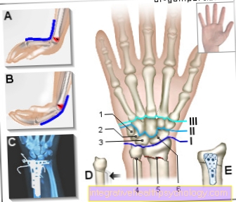
Anatomy of the talus
The center of the talus is called the Corpus tali designated. The corpus lies cranially (above) Trochlea tali (Joint roller). It stands over a cartilaginous, curved joint surface with the underside of the Tibia (Tibia), the roller roof (Facies artiucularis inferioir tibiae), and forms the distal (further away, further down) joint body of the Upper ankle (OSG).
Behind the trochlea tali, the talus has a bony protrusion, the Processus posterior tali.
The corpus tali passes forward via the collum tali (neck) into the caput tali (head). The caput tali is involved in the formation of the cartilaginous joint surface lower ankle (USG) involved.
Downwards the corpus tali forms three articulations with the Calcaneus (Heel bone).
Appointment with ?

I would be happy to advise you!
Who am I?
My name is dr. Nicolas Gumpert. I am a specialist in orthopedics and the founder of .
Various television programs and print media report regularly about my work. On HR television you can see me every 6 weeks live on "Hallo Hessen".
But now enough is indicated ;-)
Athletes (joggers, soccer players, etc.) are particularly often affected by diseases of the foot. In some cases, the cause of the foot discomfort cannot be identified at first.
Therefore, the treatment of the foot (e.g. Achilles tendonitis, heel spurs, etc.) requires a lot of experience.
I focus on a wide variety of foot diseases.
The aim of every treatment is treatment without surgery with a complete recovery of performance.
Which therapy achieves the best results in the long term can only be determined after looking at all of the information (Examination, X-ray, ultrasound, MRI, etc.) be assessed.
You can find me in:
- Lumedis - your orthopedic surgeon
Kaiserstrasse 14
60311 Frankfurt am Main
Directly to the online appointment arrangement
Unfortunately, it is currently only possible to make an appointment with private health insurers. I hope for your understanding!
Further information about myself can be found at Dr. Nicolas Gumpert
causes
A Talar fracture requirement strong violenceas they e.g. occurs in a traffic accident or a fall from a great height - i.e. when strong forces act on the ankle joint and the talus. The fracture of the Processus lateralis tali is increasing at Snowboarders diagnosed, from where the name of this talus fracture comes from: Snowboarders ankle.
Diagnosis
An important clue for the doctor lies in the anamnese, the description of the situation in which the injury occurred. Continue to be on Mobility of the foot (motor skills) and whether Loss of sensitivity (the sensation in and on the foot).
X-rays in several planes (lateral and anterior-posterior) can provide more precise information about the talus fracture. Further diagnostics can also be carried out in the Computed tomograph (CT) should be appropriate to show a possible fracture more precisely.
As further measures in the Aftercare can do that MRI and the Bone scintigraphy be a decisive means to exclude or detect possible damage in the affected bone parts.
Frequency distribution
The talar fracture is more of one of the rarer fractures. It accounts for less than 5% of all fractures in the foot area. A talar fracture often arises in combination with other fractures in the foot area, e.g. of the Malleoli (Ankle) or des Calcaneus (Heel bone).
In half of all cases of a talus fracture, the neck of the talus is affected. Fractures of the ankle bone account for about a quarter, while fractures of the bony protrusions of the talus (processus) make up about a fifth of all cases.
Symptoms:
If there is a talar fracture, exist strong pain in the field of Ankle joints. In addition, a severe swelling With Hematoma (Bruise). In addition, the Mobility in the hocks limited.
Classification
The classification a talus fracture at the talus neck is performed according to Hawkins guidelines.
At Type 1 finds no shift of the Collum tali.
Type 2 is present when the broken Talus neck in the lower ankle moves forward.
Type 3 describes a state in which the Ankle body in the upper and lower ankle postponed is.
At Type 4 the status is as with type 3 and additionally one shift in the articulatio talonavicluare. The talonavicular joint is the joint between the caput tali and the os naviculare (scaphoid bone).
therapy

A talus fracture with displacement should be reduced as quickly as possible (bring it back into the correct position) to reduce the risk of osteonecrosis (Death of the bone) to be kept as low as possible.
In the case of fractures of the lateral and posterior tali processes, a distinction is made between displaced and non-displaced fractures. If there is a dislocation of the bone fragments, then these will be with Screws moved back into position. If the fracture does not show any displacement, treatment with one is sufficient plaster castwho that Ankle immobilized (immobilized).
Talar fractures on the talar head are usually fixed with screws. Here is one Cancellous surgery can be indexed. This is a process in which healthy bone tissue (spongosia) of the patient is introduced into the fracture with the aim of growing together with the bone tissue there stable bone substance let develop. The healthy bone tissue is usually taken from bone that is easily accessible (e.g. parts of the iliac crest).
In the case of fractures of the Collum Tali can also be used plaster be worked if there is no dislocation of the fracture pieces. If a dislocation has occurred, as in Hawkins 3 and 4, and often also in Hawkins 2, one will occur Restoration with screws instead of.
If with a fracture small fragments of bone if what often happens on the small bony protrusions that are not held by screws and therefore cannot be reduced, then there is the possibility of arthroscopic removal these very fragments.
A complete load on the affected foot should be prevented up to the 8th to 12th week. A Partial load can also be possible beforehand if screws were used.
After the operation is a radiological follow-up the fracture is indicated in order to control the healing process and identify complications. Lymphatic drainage and physical therapy can be helpful to speed up the healing process.
Complications
The Blood supply of the talus occurs through several small vessels lying in a confined space. Dislocations can easily injure them. This is one of the reasons why with a talar fracture risk for osteonecrosis (Death of the bone) is very high. With Hawkins I, the risk is around 10%, while with Hawkins 4, a risk rate of 80-100% is spoken of.
Further complications lie in the development of Osteoarthritis in the upper and lower Ankle joint. Because the talus consists largely of articular surfaces, there is a likelihood of a Articular surface and thus Cartilage damaged becomes, very high. On the damaged one cartilage the wear and tear in the joint is significantly increased and the development of a arthrosis more likely.
Furthermore, in the course of a talus fracture, a Tarsal tunnel syndrome arise. This is a bottleneck that compresses nerves running in the foot and restricts their function - this often manifests itself in one Numbness on the soles of the feet.