Therapy of esophageal cancer
All information given here is only of a general nature, tumor therapy always belongs in the hands of an experienced oncologist!
Synonym
Esophageal Carcinoma, Esophageal Tumor, Esophageal Tumor, Esophagus-Ca, Beret Carcinoma
definition
The esophageal cancer (esophagus = esophagus) is a malignant, uncontrollably fast growing tumor that originates from the cells of the esophageal mucosa.
In 80-90% of cases there is a connection between long-term consumption of high-proof alcohol (alcohol abuse) and the consumption of cigarettes. The esophageal carcinoma can also develop from a beret esophagus, which is a consequence of reflux disease (chronic heartburn). The tumor causes symptoms late, when it is already well advanced. Due to the late diagnosis, this type of cancer has a very poor prognosis for patients.
Tumor stage

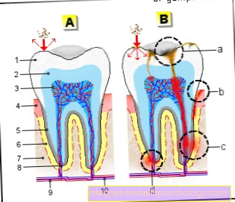
Illustration esophageal cancer
The tumor has already closed a large proportion of the esophageal diameter. This results in difficulty swallowing.
The tumor stage is now determined by the diagnostics mentioned above. The tumor stage is decisive for further therapy planning. However, an exact assessment of the tumor stage is often only possible after the operation.
To determine the tumor stage, the TNM classification used:
The T stands for the tumor size and its extent in the wall layers.
The N stands for the number of affected lymph nodes
The M. for tumor settlements (metastases) in other distant organs.
All procedures are exemplary, the concrete procedure must always be made dependent on the individual case of the patient.
Stage 0 = carcinoma in situ
The cancer is only in the top layer of cells and has no connection to it Lymphatic system.
The tumor is surgically removed (Mucosal resection).
Stage I.
The tumor is limited to a small part of the esophagus. It has not spread to neighboring tissue, lymph nodes or even other organs.
The surgery involves removing part of the esophagus, in some cases the entire esophagus (Esophagectomy). The lymph nodes are also removed. Radiochemotherapy follows after the operation.
Stage II
The cancer has taken up large parts of the esophagus. Regional lymph nodes may already be affected, but no other organs and tissues.
Often treated like stage I.
Stage III
The cancer has spread to tissues or lymph nodes near the esophagus. However, it does not yet affect any distant organs.
The operation tries to remove the tumor with the aim of relieving pain and discomfort. If the tumor is difficult to operate, chemotherapy and radiation (chemoradiotherapy) will be performed prior to surgery to shrink the tumor so that it is easier to operate (neoadjuvant therapy). With this therapy you can lower the tumor stage (so-called Downstaging).
Stage IV
Cancer cells have already spread to other parts of the body and organs.
First, an irradiation is carried out to reduce the tumor mass. Sometimes filing is one Stents into the esophagus. This stent is a type of tube that holds the esophagus open. Part of the tumor can be removed using a laser or electricity (electroresection).
therapy
The treatment of the patient requires intensive cooperation between the medical departments of surgery, internal medicine and radiation therapy.
In therapy, the TNM classification is used as an essential aid to decision-making. There are corresponding therapy guidelines for each tumor stage. So one can describe three treatment goals that are considered depending on the stage.
Healing of the patient
This goal exists for patients in stage I (see above). A recurrence of the carcinoma in the same place is theoretically possible (tumor recurrence) if individual tumor cells should have survived the operation and chemoradiation. Careful follow-up is therefore necessary.
Tumor control
In patients with tumor stages II and III, various therapeutic approaches are used to control tumor growth and prevent it from spreading. Despite the lack of healing options, a long-term freedom from symptoms is sometimes achieved.
Symptom relief
Cure is impossible in patients with stage IV tumors. The main focus of the therapy is to alleviate symptoms (especially pain, difficulty swallowing, eating).
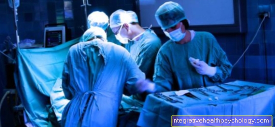
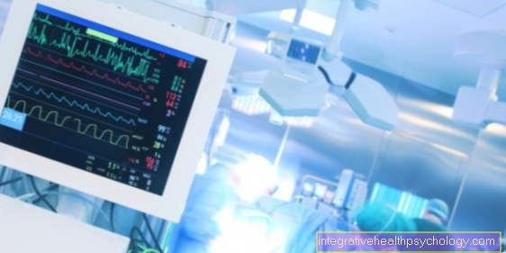
Operative therapy
If the tumor is only in the superficial layer of the esophageal mucosa, it can be removed superficially during an endoscopy (mucosal resection).
In a transthoracic esophagectomy, both the chest (thorax) and the abdomen (abdomen) are opened and the affected esophageal section is removed with sufficient distance to the tumor and the surrounding lymph nodes.
In some cases, the entire esophagus must be removed. If the lower esophagus or the junction with the stomach is affected, the stomach must also be partially or completely removed. Sometimes it is necessary to remove structures in the vicinity as well as the surrounding fatty and connective tissue. The removed section of the esophagus is either replaced by the stomach that has been moved into the chest (stomach pull-up) or, if the stomach has also been removed, by a piece of the intestine.
Pathological diagnostics
The removed esophageal tumor is assessed histologically after removal. For this purpose, the tumor is cut at certain points and some samples are taken. From these samples, wafer-thin sections are made, stained and assessed under the microscope. The tumor type is determined and the lymph nodes with removed are examined for tumor involvement. In order to completely rule out lymph node involvement, the pathologist must examine at least 6 lymph nodes. The tumor can only be clearly described according to the TNM classification after the pathological findings.
radiotherapy
In most cases, the esophageal cancer responds well to radiation therapy.
In some cases, radiation therapy is first used before the operation (neoadjuvant) to reduce the tumor and thereby make it operable. If radiation therapy is initiated after surgery (adjuvant), the risk of a local tumor recurrence (recurrence) can be reduced.
Even patients who have little prospect of a cure are sometimes subjected to radiation therapy (palliative radiation). Radiation therapy can reduce the tumor mass and thus alleviate tumor pain and difficulty swallowing.
The radiation therapy can be carried out externally, i.e. through the skin (percutaneous). In the case of tumors that constrict the esophagus, small-area irradiation (brachytherapy) can be carried out from within (intraluminal afterloading). With this type of radiation therapy, a tube is pushed into the esophagus and a small radioactive source of radiation is introduced through it. In this way, the tumor can be irradiated from within.
chemotherapy
The esophageal cancer only responds moderately to chemotherapy, so that in most cases you get the radiotherapy and the chemotherapy combined (Chemoradiotherapy) in order to potentiate the therapeutic benefit. For the treatment of this type of cancer, the Cytostatics 5-fluorouracil, cisplatin and Taxanes administered.
Stent insert
To keep the esophagus open for food to pass through, a plastic tube (stent) must occasionally be inserted into the esophagus.
Stents are also used after a partial esophageal removal operation. Here, the connection point between the remains of the esophagus and the raised section of the intestine, the so-called anastomosis, can be sealed and stabilized with a stent.
Laser therapy
If you can no longer operate on the tumor and food intake is severely impaired, laser therapy can provide symptom relief. Here, parts of the tumor are vaporized by the laser, which reduces the extent of the esophageal narrowing and allows food to pass through the esophagus more easily. Unfortunately, the tumor often grows back from the lower layers, so that the treatment sometimes has to be repeated after 7-14 days.
If laser therapy is combined with small-area radiation (brachytherapy), the time until re-treatment can be significantly extended.
In tumor stage 0, in which only the top layer of cells in the esophagus is affected, laser treatment can also be used to remove the tumor.
Photodynamic Therapy
The photodynamic therapy is still being tested and is only used in patients with a small tumor without lymph node metastases.
In this therapy, the patient is initially given a drug (so-called porphyrins) administered, which make the tissue extremely light-sensitive. Three days after drug administration, the tumor is irradiated with laser light so that it can be destroyed due to the artificially generated sensitivity to light.
Note photodynamic therapy
The patient should be particularly careful to protect his skin from sunlight during the therapy period.
Nutritional fistula
If other therapy options to keep the esophagus open fail, a feeding tube (PEG; Percutaneous Endoscopic Gastrostomy) can be placed directly through the skin into the stomach. This method of treatment is a small surgical procedure. Under endoscopic control, a hollow needle (cannula) is first pushed through the skin and into the stomach in order to insert a plastic tube over it as a permanent connection to the stomach.
The PEG offers many advantages for the patient in contrast to a gastric tube inserted through the nose. The patient can feed himself through this tube. The tube clogs less easily than the nasogastric tube and you can feed more food at once.
The most important point for the patient, however, is aesthetics, as the tube disappears under clothing, invisible to others.
forecast
The Esophageal carcinoma Overall, esophageal cancer has a poor prognosis because most esophageal tumors are discovered at a late stage. Five years after the initial diagnosis was made, only 15% of all tumor patients are still alive.
The tumors further down the esophagus have a slightly better prognosis. The further up (near the mouth) the tumor sits, the worse the prognosis.
On average, tumor patients only live 8 months after the first occurrence of a swallowing disorder.