Aortic stenosis
What is aortic stenosis?
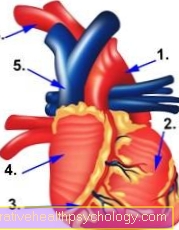
Aortic stenosis is the short form of aortic valve stenosis and describes a congenital or acquired heart valve disease. In aortic stenosis, the aortic valve, the heart valve between the left ventricle and the main artery, is pathologically narrower than in healthy people. A progressive calcification of the valve pockets of the aortic valve, which stiffen the tissue of the heart valve more and more and make it more immobile, is typical.
In the case of a stenosis, the valve is so abnormally changed that it no longer opens completely. As a result, blood flow through the heart valve into the main artery (aorta) is impaired. A progressive stenosis of the aortic valve can have serious consequences and must therefore be clarified by a doctor.

The reasons
Aortic stenosis can be congenital or develop in the course of life (acquired aortic valve stenosis). Most aortic stenoses are acquired and are a common disease in old age. It is also known as denegerative aortic valve stenosis. Age-related stenosis is mainly caused by wear processes such as calcification.
An unhealthy lifestyle, which causes permanently high blood lipid levels through fatty food and lots of meat, promotes the development of aortic valve stenosis. Rheumatic fever and endocarditis can leave scar tissue on the aortic valve, causing stenosis. The congenital form of aortic stenosis is hereditary.
Find out all about the topic here: The aortic stenosis.
The symptoms
If the aortic stenosis is low, there are usually no symptoms. If the stenosis becomes worse, those affected often complain of dizziness, and occasionally they suffer from fainting (syncope). Symptoms of dizziness and fainting are due to insufficient blood flow to the brain through the aortic stenosis.
With pronounced stenosis of the aortic valve, severe symptoms such as angina pectoris (chest tightness) and dyspnoea (shortness of breath) occur. Angina pectoris is an attack-like pain in the chest that is caused by a short-term reduced blood flow to the heart. Chest pain can vary in severity and can feel dull, stabbing, burning, or heavy. The tightness of the chest is often accompanied by a feeling of suffocation. Dyspnea is the subjective feeling of not getting enough air. Those affected then breathe in more often, so that the breathing rate increases significantly. Angina pectoris and shortness of breath are generally very unspecific symptoms for which many cardiovascular diseases are possible.
A cardiac exam with thorough auscultation is important to make the correct diagnosis. Patients with pronounced aortic valve stenosis often feel weak and tired.
Find out more about the topic here: Angina pectoris.
The diagnosis
A thorough examination is necessary to make the diagnosis of aortic stenosis. During the clinical examination, the cardiologist often notices a low amplitude of blood pressure with a sluggish increase in pulse rate. A heartbeat can be palpable. It is important to listen to the heart (auscultation), during which a typical heart murmur can be heard.
Further examinations are usually carried out to confirm the diagnosis. A chest x-ray provides information about an enlarged heart (due to the increased work of the heart) and calcification of the aortic valve. Typical signs of aortic stenosis can be read in the ECG (electrocardiogram) and heart valves and heart function can be assessed very well in a cardiac ultrasound examination (echocardiography). In addition, a cardiac catheter examination can be carried out, which can also be used as an invasive therapy method.
Read more about the topic here: The cardiac catheter examination.
The auscultation
Auscultation is an important diagnostic tool that can corroborate suspicion of aortic valve stenosis. Auscultate over the chest with a stethoscope. You can listen to specific points where the tones and noises of the various heart valves are projected.
With aortic valve stenosis, there is a characteristic heart murmur that can be heard loudest between the second and third ribs to the right of the sternum. In cardiology, the heart murmur is described as a spindle-shaped, rough systolic which radiates into the carotid arteries and the maximum punctum in the second intercostal space to the right of the sternum (parasternal). Experts may hear an “early systolic ejection click” and, if the aortic valve is immobile, a quieter second heart sound. If the aortic stenosis occurs together with an aortic valve insufficiency, there is a so-called early diastolic decrescendo.
As a non-medical practitioner, one can remember that the cardiologist, when listening to the chest, often hears a noise above the aortic valve that is specific to a narrowing of the heart valve. A corresponding heart murmur reinforces the suspicion of aortic valve stenosis and usually requires further diagnostics.
The heart echo
A heart echo (echocardiography, heart ultrasound) is an examination that is performed with an ultrasound device over the chest (transthoracic echocardiography) or over the esophagus (transesophageal echocardiography). It is a method that can be used to thoroughly analyze the heart valves and detect valve defects such as aortic valve stenosis
The echocardiography findings are also used to evaluate the severity of the stenosis.
Find out more about the topic here: Echocardiography.
The introduction
Aortic valve stenoses are initially divided according to their origin, i.e. acquired or congenital (inherited). In inherited aortic stenosis, the localizations of the narrowing on the aortic valve must be distinguished: valvular / supravalvular / subvalvular aortic stenosis. The shape of the aortic valve can be unicuspid or bicuspid and refers to the presence of certain heart valve structures.
With the aid of the heart ultrasound examination, the mean pressure gradient of the aortic valve, the aortic valve opening area and the “valvular restiance” (valve resistance) are determined. These criteria are used to assess the severity of aortic valve stenosis. The degrees of severity are graded as mild, moderate and high
The treatment
The treatment of aortic valve stenosis depends on the severity of the stenosis. If there is a slight narrowing of the aortic valve, treatment is usually conservative at first. In the case of aortic stenosis, this means doing without heavy physical exertion, and those affected should take it easy. Endocarditis prophylaxis is also recommended to prevent inflammation of the heart valve. This includes, for example, the administration of antibiotics during (surgical) interventions so that pathogens on the heart valves have no chance.
If the aortic stenosis is more pronounced and clinical symptoms are present, surgical therapy is preferred. There are several surgical methods to treat stenosis. For acquired stenosis, which often occurs in old age, an aortic valve replacement is often chosen. There are biological valve prostheses from pigs, cattle or horses, and mechanical valve prostheses. Valves from human corpse donors are rarely used. An alternative is balloon dilatation of the aortic valve. This method is mainly used for congenital aortic stenosis and is performed with a cardiac catheter.
Ross surgery is also performed on young patients with congenital aortic stenosis. In this method, another heart valve (the pulmonary valve) is replaced by a third party and your own pulmonary valve is used as the new aortic valve. The advantage is that this heart valve grows very well with young patients.
For more information, read on: The artificial heart valve.
The drugs
People with aortic stenosis and heart failure for which surgery is not an option are given diuretics and possibly digitalis glycosides. The use of diuretics should be done carefully and initially in low doses.
There are also drugs that are absolutely contraindicated for aortic stenosis and must not be administered. These include the ACE inhibitors, which lower the so-called afterload of the heart. In the case of aortic stenosis, these agents would additionally increase the pressure and are therefore strictly prohibited. Calcium antagonists and nitrates are also taboo.
Also read the article: The diuretics.
When do you need an operation?
Surgery is an option for aortic stenosis as soon as symptoms appear. Clinical symptoms often occur with moderate to severe stenoses. There are various options for those affected to treat the aortic valve surgically.
How dangerous is surgery?
Operations involve risks and dangers, but these basically depend on the respective method and the requirements of the patient. Additional heart diseases, anesthetic tolerance and other factors play an important role.
Open heart valve operations are an option for “fit” patients. For patients for whom open surgery seems too risky, balloon dilatation with a cardiac catheter can be a suitable procedure.
The course of the disease
Untreated aortic valve stenosis usually worsens the stenosis. If the cause is valve wear and an unhealthy lifestyle, calcification will progress and the valve will tighten. If left untreated, dangerous complications can arise.
The turbulent blood flow on the damaged heart valve can cause small blood clots to form, be carried away with the bloodstream and transported to the brain. There they can clog a vessel and cause a stroke. Untreated aortic stenosis can also cause dangerous cardiac arrhythmias and lead to cardiac death via ventricular fibrillation.
However, if a stenosed aortic valve is treated surgically, the prognosis is good and the course of the disease is positive.
Does aortic stenosis limit life expectancy?
With aortic stenosis, life expectancy depends largely on the severity of the vascular narrowing. Comorbidities and the general condition of the person affected are important factors.
The earlier an aortic stenosis is detected, the better the prognosis. With untreated stenosis, life expectancy is severely reduced over time and dangerous complications can occur, including death. If aortic valve stenosis is successfully operated on, the prognosis is good these days.