Femoral artery
General
The arteria femoralis (large leg artery), arises in the pelvis from the external pelvic artery (External iliac artery). It is then initially between the nerve and the vein (Femoral nerve and Femoral vein) and can be easily felt at this point in the area of the inguinal canal.
For this reason, the femoral artery is often used for puncture during cardiac catheter examinations or to insert a central catheter.
The femoral artery supplies the thigh with oxygen-rich and nutrient-rich blood. Since the muscles of the thigh are the largest muscle group in the body, they need a particularly good blood supply.
Read more on the subject at: Thigh muscles
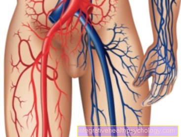
Location and course
Below the Inguinal ligament (Inguinal ligament) the artery runs on one of the Pelvic bones (Pecten ossis pubis) and moves from there to the Trigonum femoral (Iliopectineae fossa), which by the Iliopsoas muscle and the Pectineus muscle is limited.
From there the artery closes Back of thighs. On her way there she runs together with the Saphenous nerve through the Adductor canal. At the exit of this channel goes the Femoral artery in the Popliteal artery above. This way branch different vessels from the femoral artery.
Superficial femoral artery
The Main branch after the branching off of the deep femoral artery, the femoral artery is also known as the superficial femoral artery (superficialis Latin for "superficial"), as this superficial in the skin located distally and finally in the hollow of the knee Popliteal artery transforms. The vessel is covered by the Fascia lata between the iliopsoas and pectineus muscles from the groin to the hollow of the knee. The artery passes through other structures, such as the Adductor canalwhich it leaves through the adductor hiatus and is then referred to as the popliteal artery.
The branches of the superficial femoral artery are the superficial epigastric artery, the superficial circumflex ilium artery, the pudendal artery and the deep femoral artery.
Thus, the superficial femoral artery supplies part of the skin of the abdominal wall, the external genital organs, the knee and parts of the lower leg, then already known as the popliteal artery.
Deep femoral artery
The deep femoral artery (profunda lat. for "deep") is that greatest finish the arteria femoralis, which subsequently also bears the name Arteria femoralis superficialis, and runs in the deep of the thigh. It is mainly responsible for the supply of the thigh and gives off several branches.
The important branches of the deep femoral artery are the A. circumflexa femoris medialis and lateralisthat come into contact with one another in the trochanteric fossa on the thigh and form an anastomosis. For the back of the thighs, branch Arteriae perforantes from.
More branches
The Superficial epigastric artery branches directly in the Inguinal canal from the femoral artery and from there upwards again towards the trunk.
The different External pudendal arteries take care of the woman labia minora and with the man Scrotum, as well as in both sexes the Groin skin with arterial blood.
Another small branch is that Superficial circumflex iliac artery. This artery serves to supply part of the Ilium.
The artery lying on the inside, the arteria circumflexa femoris medialis, serves the Supply of ischiocrural Musculature, the lateral circumflexa femoris artery supplies the Extensor muscles (Extensors) of the thigh.
On the other hand there will be three to four Arteriae perforantes released, which reach the back of the thigh and supply it with oxygenated blood. The inside of the thigh is through the Genicular descending artery supplied, which together with the Saphenous nerve through a small gap in the muscle layer that Septum intermusculare vastoadductorium, pulls.
How can I feel the femoral artery?
The palpable pulse of the femoral artery is called the femoral pulse. It can be felt in the groin area. Several fingers should be used at the same time to feel the pulse. The thumb should not be used. While the key is being pressed, the elapsed time should be determined with the aid of a clock in order to be able to calculate the number of beats per minute. To find the femoral pulse, first palpate the pelvic bone from the front of the body. On each side of the body there should be a small protrusion of bone a little below the level of the navel. From this palpable point, an imaginary line is drawn to the so-called symphysis. This is the palpable connection point of the pelvic bones in the middle of the body. It lies just above the genital region. The femoral pulse should be palpable in the middle between the two points.
Stenosis of the femoral artery
Stenoses of arteries can be divided into thrombosis and embolism:
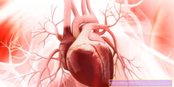
If a blood clot is washed into the arterial vessels of the leg, e.g. coming from the left heart, it is an acute occlusion, an embolism.
If a blood clot forms on the vessel wall as a result of an atherosclerotic disease, stenosis of the artery can occur. It's an ongoing process, a thrombosis.
The vessel is then partially or even completely closed, so that the tissue of the extremity behind it can no longer be adequately supplied with oxygen and nutrients. There is ischemia of the supply area, which is reflected in the pallor and coldness of the skin and severe pain. If the occlusion remains for a long time, this leads to tissue necrosis and even gangrene, i.e. the complete death of the affected area.
Read more on the subject at: Thigh pain
Typical of a stenosis of the femoral artery is pain that is exerted by exertion and disappears again after the exertion has ceased.
The aim of therapy is reperfusion of the ischemic area in order to prevent necrosis. In an acute emergency or in the case of very advanced occlusions, an operative intervention (e.g. a bypass) is carried out to restore the blood flow to the relocated supply area. If the occlusion of the artery is not very advanced, changes in lifestyle (no smoking, change in diet, etc.) will help to improve the condition and prevent further enlargement of the thrombus.
Treating a stenosis with the stent
The femoral artery can narrow over the course of life, making it very difficult for blood to flow into the leg. The result is pain in the leg, especially under stress. Treatment can be done in different ways. A so-called stent is used especially in severe cases. This is a small tube made of mesh-like wire mesh. It usually consists of synthetic fibers or special metal. The tubular braid is placed inside the artery so that it rests against the artery wall and keeps it open. The examiner may widen the artery beforehand. The inserted stent stabilizes and supports the artery. The stent is implanted in a minimally invasive manner using a so-called catheter. All that is needed is a small opening in the vessel through which the stent and catheter can be inserted.
Read more on the subject: Stent
Femoral artery aneurysm
In the superficial and deep femoral arteries, damage to the intima of the vascular wall, i.e. the innermost layer, can develop Aneurysm come. This leads to a bulging of the vessel wall. With a certain form of the aneurysm, the parts of the vessel wall, intima and media, separate from each other due to a bleeding that continues to proliferate. The Bleeding can cause a wrong one second vessel opening, a so-called pseudo-lumen, arises within the vessel wall. At the same time, the normal lumen of the artery is narrowed.
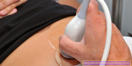
Aneurysms can congenital or acquired be and are mostly symptom-free in the early stages, so that only one Incidental finding get discovered.
Possible consequences of aneurysms are on the one hand Blood clotswhich can form on the defective vessel wall and thus lead to a further narrowing of the vessel or are transported further into vessels lying further on the periphery and one complete closure cause.
On the other hand, there is a risk of Rupture of the aneurysm, i.e. the tearing of the sac, with potentially dangerous bleeding.
Depending on the condition of the aneurysm at the time of diagnosis, surgical intervention can be the treatment of choice here as well.