biopsy
Definition - what is a biopsy?
Biopsy refers to the removal of tissue, the so-called "biopsy", from the human body in clinical diagnostics. It is used to examine the cell structures removed under the microscope. In this way, initial suspected diagnoses for potential diseases can be safely confirmed.
The doctor in charge carries out the biopsy in different ways. A needle is pierced from the outside into the tissue to be examined in order to obtain a tissue sample.
The most common type of biopsy is a fine needle biopsy. It is mainly used to obtain cells from internal organs and tumors. Although the method is very gentle and painless, several thousand cells can be obtained using a slight negative pressure.
The fine needle biopsy is traditionally used for thyroid biopsy.
Other options for biopsy include curettage (scraping the uterus after a miscarriage), punch biopsy, incisional biopsy and vacuum biopsy. In addition to these, there are numerous other techniques for performing a biopsy. An invasive biopsy is also possible, in which a skin incision is made beforehand in order to make the area to be examined more accessible.
Appraisal
The word biopsy translated from Greek means: See life (Bios = life; Opsis = seeing). It delivers one Possibility of safe diagnosis after a suspected clinical diagnosis. After performing the actual biopsy, a pathologist receives the tissue samples. The pathologist examines the cells under the microscope and can then make statements as to whether the tissue is healthy or abnormally changed. This one The field of medicine is called "pathohistology".
A biopsy is meaningful for many diseases of the internal organs, especially if there is a suspicion of tumor diseases. Only a biopsy can reliably determine whether the tumor is benign or malignant. On the basis of the fine-tissue cell structures, the pathologist not only recognizes whether the cells of the organ are healthy or not, but also what forms of change are involved and which organ they originally come from. Particularly in the case of metastases from malignant tumors in other organs, the original tumor can be determined by a biopsy.
What types of biopsy are there?
There are a number of different types of biopsy. The most common types of biopsy include:
- Incisional biopsy
- Excisional biopsy
- Biopsy punch or punch biopsy
- Fine needle biopsy
- Suction biopsy or vacuum biopsy.
A distinction is made between open biopsy forms (sample excision) and minimally invasive forms of biopsy. Incisional and excisional biopsies are among the open forms of biopsy. The minimally invasive forms of biopsy include the punch biopsy, the fine needle biopsy and the suction biopsy.
The incisional biopsy refers to the removal of part of a tissue change, while an excisional biopsy is the complete removal of a tissue change and a small part of the surrounding tissue.
With the biopsy punch, punch cylinders are removed from suspicious tissue using a special device. It is often used for biopsies of the mammary gland and prostate. In a fine needle biopsy, a fine cannula (hollow needle) is punctured through the skin and the tissue sample (biopsy) is removed using a negative pressure created by a syringe attached.
The suction biopsy is performed using a special needle, which consists of an outer and an inner needle. Computer-controlled, this is guided to the destination and the tissue sample is removed.
Imaging methods such as ultrasound or computer tomography are often used to support the various forms of biopsy. This increases the likelihood that the biopsy will contain a sample from the suspect area.
Incisional biopsy
With an incisional biopsy, only part of a suspicious tissue is removed. This type of biopsy has a very high accuracy, as sufficient characteristic tissue is removed compared to the other types of biopsy. Depending on where the incisional biopsy is to be performed, either a local or a short anesthetic is given. The disadvantage is that there is a higher risk of bruising (hematoma) compared to the other types of biopsy.
Biopsy punch
A biopsy punch, also known as a punch biopsy, is performed using a special device. This is often performed using ultrasound or X-ray control in order to achieve a high level of accuracy and to minimize risks such as injuring neighboring structures. It is mainly used for biopsies of the mammary gland and prostate, but can also be used for liver biopsies, for example. The biopsy punch removes tissue cylinders from the suspect tissue. A pathologist then examines the tissue (histologically).
Fine needle biopsy
A fine needle biopsy is used to obtain cells from internal organs. It is performed using a thin needle with a hollow channel in the middle. It is mainly used to puncture lung tissue or bone marrow. Individual cells are obtained. These are sucked in by means of a negative pressure created by an attached syringe.
It has the advantage that the complication rate is very low. The risks are lower and a potential spread of the tissue (e.g. tumor cells) is minimized.
The disadvantage is that the fine tissue assessment is quite difficult, since only little material is obtained. If there is any uncertainty, another biopsy may have to be performed.
Vacuum biopsy
A vacuum biopsy, also known as suction biopsy, is usually only carried out if the biopsy cannot be clarified using a sonographic punch biopsy. It is mainly used for biopsies of the mammary gland and the prostate. It is characterized by a high level of accuracy. This means that the tissue which is obtained has a high probability of retaining part of the suspicious tissue. Usually several pieces of tissue are removed to increase accuracy.
In a vacuum biopsy, the biopsy needle consists of an outer and an inner needle. Before the biopsy, a small skin incision is made through which the biopsy needle is passed. The biopsy needle cuts a small piece of tissue from the suspicious area. The piece of tissue is then sucked into the extraction chamber of the outer needle by the vacuum generated. Like all biopsies, the tissue is examined by a pathologist.
How does a biopsy needle work?
Biopsy needles are available in different lengths and with different inner diameters. A biopsy needle is a hollow needle. If a syringe is placed on a biopsy needle, a negative pressure can be generated. The tissue cylinder can be sucked through this and sucked into the interior of the needle. This is called aspiration. Today, most biopsy needles are fully or semi-automatic.
There are also special needles, such as in the vacuum biopsy, which consist of an outer and an inner needle.
What are the risks of a biopsy?
Risks with a biopsy can be bleeding and bruising at the donation site. They are more common than the rest of the risks. The risk of bleeding is increased if organs with good blood flow are biopsied or blood-thinning medication is taken.
It is also possible that neighboring organs or structures are injured. The risk can be minimized by using imaging methods such as ultrasound or X-rays.
Further risks can be wound infections or wound healing disorders. However, these only occur very rarely.
It is currently being discussed whether tumor cells can be carried over by a biopsy and whether metastases can form in the extraction channel as a result. However, this is described as very unlikely in the current literature.
Breast biopsy
Due to the constant remodeling of breast tissue in women, there is a permanently increased risk of tissue changes. Most women discover knot-like structures on their own breast in the course of their lives, which require further clarification. In most cases, these are benign nodes. Theoretically, however, a malignant tumor can be present and should be treated as soon as possible.
After a suspected diagnosis has been made, a biopsy is taken from the breast tissue. A high-speed punch biopsy is usually performed for this purpose. For this purpose, the suspicious tissue is punched three times under control with an ultrasound device. This happens at such a high speed that the pain is very little. All that is needed beforehand is a local anesthetic and a small skin incision. The risk of bleeding and infection is very small. With fast punching processes, there is always a small risk of spreading tumor cells that can settle in another location and spread again (metastases).
The punch biopsy in the diagnosis of breast tumors is a popular procedure. Your results can be classified as very meaningful. If at least 3 biopsies are carried out, there is a high degree of certainty that a sufficient number of abnormal cells will be obtained. Benign tissue is recognized as such with great certainty; the diagnosis of malignant tumors is true with a probability of 98%. In many cases, it saves women from hasty surgical interventions due to misdiagnosis after previous mammograms.
Other biopsy methods that can be used on the breast are fine needle biopsy, extirpation, mammotomes and other punching methods.
Read more about breast cancer tissue samples and here Breast biopsy
Biopsy of the cervix
The biopsy of the cervix is known as a colposcopy-guided biopsy. Colposcopy is a gynecological examination in which the vagina and cervix can be examined with the help of a special microscope. At this stage, a biopsy of the cervix can be performed if tumorous changes are suspected. Tissue samples are taken from the suspicious areas with small forceps (snap biopsy) and then forwarded to the pathologist for a histological examination. Usually this is not painful.
Read more on the subject at: Cervical biopsy
Prostate biopsy
Prostate cancer is a very common type of cancer in men of advanced age. As part of preventive programs, every man over the age of 45 should have an annual prostate examination. This consists of a palpation examination of the prostate and a determination of the PSA level in the blood.
Read more about prostate cancer and prostate examinations here
A conspicuous examination finding, for example an enlarged prostate associated with an increased PSA value, always suggests prostate cancer and should be examined further. A benign enlargement of the prostate, a so-called "benign prostatic hyperplasia", or an inflammation (prostatitis), can increase the PSA level in the blood.
Read more about inflammation of the prostate here
A prostate biopsy may be necessary to make a reliable diagnosis. It is an uncomplicated and safe procedure and its results are meaningful. So far, the biopsy is the only way to make the definitive diagnosis of prostate cancer.
Here you can find more information about prostate biopsy.
After a thorough explanation by the doctor, the biopsy can be performed in the hospital on an outpatient or inpatient basis. It should be noted that the biopsy as an invasive procedure is always associated with risks. In addition to infection and bleeding, the biopsy itself can very rarely cause any tumor cells to be carried over. The subsequent results of the biopsy always include a low residual risk of being false positive (patient is healthy, but test positive) or false negative (patient sick, test negative).
During prostate biopsy, about 10 samples are usually obtained to make the procedure safer. The "prostate punch biopsy" is carried out through the anus, just like the urologist's examination. A small needle is used to numb locally and the entire procedure is controlled by ultrasound to check the correct position. Similar to the breast biopsy, the hollow needle is “shot” at high speed into the corresponding area and a punch cylinder filled with tissue is removed. After at least 10 tests, over 10,000 cells are removed from different areas of the prostate.
After the examination, there is not much to pay attention to. The procedure is largely uncomplicated. You may experience mild pain shortly thereafter, urination problems or blood in the urine.
Lung biopsy
Tissue extraction from the lungs is used relatively rarely as a diagnostic tool in the clinic. It represents an invasive, diagnostic procedure and offers the possibility to examine the lung cells histologically, immunologically or genetically for changes.
The majority of all lung diseases can already be diagnosed by the clinical appearance of the patient and subsequent radiological imaging. A lung biopsy is only necessary if the non-invasive procedure cannot reliably determine the cause of the disease. These mainly include "interstitial" lung diseases and unclear tumors. A distinction must be made as to whether the lung tissue itself, the vessels of the lungs or the lung skin, the “pleura”, is affected.
The lung biopsy can then be performed in different ways. A fine needle biopsy is also possible. It is performed without prior cuts. The needle is passed through the thorax from the outside between the ribs. The challenge here is to hit the area under investigation exactly. Ultrasound or CT can help.
Another option that is frequently used is a biopsy during a bronchoscopy. With the bronchoscope, the inside of the air-conducting airways is examined through the mouth. An integrated ultrasound head enables the infiltrate to be localized and biopsied very precisely from the inside of the bronchi.
Another very invasive method is the biopsy using thoracoscopy and thoracotomy. An incision must be made to open the chest so that samples can be taken directly from the lungs with forceps. This usually happens in the context of large, open operations.
Further information on the subject can be found at: Lung biopsy
Liver biopsy
Most of the tissue changes in the liver require a biopsy to determine the cause. This examination is usually preceded by the clinical picture and a radiological image. A liver biopsy is mainly carried out in the case of diffuse diseases of unclear origin, in the case of limited nodules that are noticeable on the radiological picture, and to diagnose genetic diseases that affect the liver, for example hemochromatosis.
The most common way to do this is a punch biopsy. Ultrasound is controlled between the ribs and a punching cylinder is removed. To keep the pain as low as possible, the patient is given a light sedative and local anesthesia at the puncture site. In other cases, biopsies can be performed as part of open or laparoscopic surgery.
When diagnosing tumor diseases, biopsies are necessary to determine the origin of the tumor or to determine whether it is a benign or malignant tumor.
Read more on the subject at: Liver biopsy
Kidney biopsy
The kidney biopsy can be performed in the case of kidney dysfunction that requires reliable diagnostic clarification.The main indication for such an examination is the “nephrotic syndrome”. This is a restriction of kidney function, which is characterized by a high excretion of proteins in the urine (proteinuria). The kidney corpuscles filter the blood, so that in the end mainly water and salts are left. Proteins are usually completely retained in the blood.
This can be caused by genetic and inflammatory kidney disease, failed transplants, or kidney failure of any cause.
The kidney biopsy is also performed using ultrasound and under local anesthesia. In some cases, the diagnosis can be made on the basis of the tissue examination of the kidney corpuscles obtained.
Read more on the subject at: Kidney biopsy
Skin biopsy
Biopsies of skin cells can also be performed and analyzed. They are carried out mainly to clarify skin findings that are visible from the outside. If the skin is abnormal, a Dermatologist Use various criteria to assess whether the change is benign or whether it requires further clarification. Depending on Appearance, size and Spread Different biopsy procedures are used to determine the findings.
In the case of smaller abnormal findings, the Excisional biopsy prefers. This is done with a scalpel the entire area cut out and then examined. With every potential result that the examination provides, the procedure on the skin is complete, as all abnormalities have been removed.
As an alternative, for example in the case of large affected areas of the skin, these are Incisional biopsy or the Punch biopsy to call. Here only one sample is obtained for examination. After the subsequent diagnosis, the remaining findings are left as they are or must be removed in a second operation.
Skin biopsies can be used with or without local anesthesia are carried out and are usually few complications.
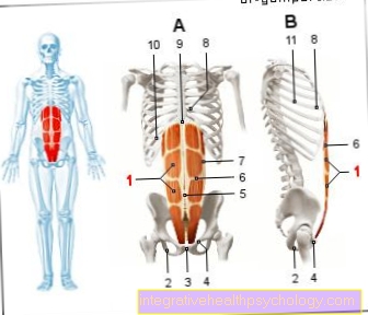
Lymph node biopsy
Lymph node biopsies are a common clinical diagnostic method, especially in cancer diagnostics. Lymph nodes may be noticed by the patient or the doctor as enlargements that can be painful. Lymph nodes can also be enlarged in the CT image. The cause can be inflammatory diseases or tumor diseases.
The lymph collects fluid from all areas of the organ and directs it back into the blood via its own lymphatic system in the neck area. With tumor diseases that spread and form settlements, so-called "metastases", the surrounding lymph nodes in particular are quickly affected. Their infestation contributes significantly to the assessment of the cancer and the decision on therapy. A particularly large number of lymph nodes are located in the groin area and in the armpit.
For an exact diagnosis, the affected lymph nodes must be biopsied. To do this, the skin is incised and the lymph node is exposed. It can then be removed and then examined cytologically and histologically. If the lymph node is actually infected with cancer, all nodes in the region are removed in order to prevent the danger of tumor cells colonizing other lymph nodes via the lymphatic system. This prophylactic intervention is known as "lymph node removal".
Read more on the subject at: Lymph node biopsy
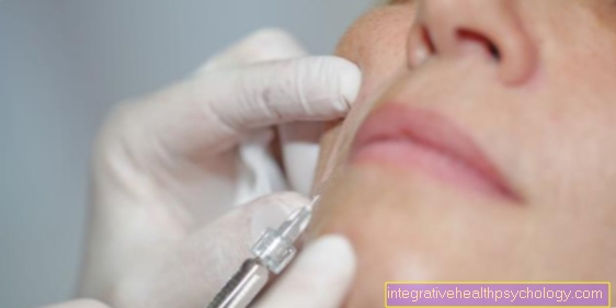
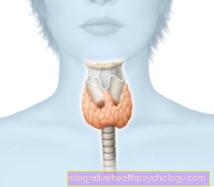
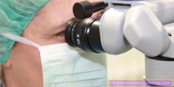
Thyroid biopsy
A thyroid biopsy is performed in clinical diagnostics for many diseases. Through previous Medical history, Scan and Ultrasound recordings of the thyroid the suspicion arises that this is abnormally changed. To the noticeable areas of the thyroid to take the biopsy by simultaneous Ultrasound recording controlled. The actual biopsy is then carried out through a fine needle. The complications fall with this method extremely low out.
May cause changes in the thyroid gland for example Inflammation be. They can arise from pathogens or as autoimmune reactions.
Also with malfunction of the thyroid gland and with Goiter formations a cause can often be found by examining the cells. In many people, the thyroid gland forms lumps, which can be active or inactive. Malignant tumors are also conceivable. Not everyone Thyroid node is in need of treatment. A biopsy is intended to provide final certainty in the event of an initially suspected diagnosis.
Biopsy of the intestine
Intestinal biopsies are common and, in contrast to many other biopsy procedures, are performed almost exclusively as part of endoscopic examinations. There are two ways of looking at the bowel, with a gastroscopy and colonoscopy. With a gastroscopy, the examination is carried out through the mouth and extends as far as the beginnings of the small intestine. With a colonoscopy, the entire large intestine and, in some cases, the end of the last section of the small intestine can be examined through the anus. In order to be able to fully observe the very long and twisted small intestine, a capsule endoscopy is necessary, in which, however, no biopsies can be performed.
With the usual colonoscopy, the biopsy sample can be obtained through the endoscope with forceps. Especially small polyps and ulcers in the intestinal wall are removed. On the basis of the samples of the mucous membrane tissue from the inside of the large intestine, inflammation, benign and malignant tumors, and other intestinal diseases can be differentiated. The biopsy in the intestine is usually not painful. During the endoscopic examination you are usually sedated and asleep. Occasionally, small amounts of blood may be found in the stool afterwards. Infection of the biopsy site is a very rare complication.
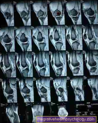
Brain biopsy
A biopsy in the brain is only carried out according to exact previous radiological examinations. Fall changes in one CT or MRI scan of the brain on, it must be assessed how quickly the structures are growing. There is no time and the change in the brain is already taking place symptomatic noticeable, a biopsy must be performed in order to be able to start therapy as soon as possible.
Such altered structures in the brain tissue can be caused by inflammatory lesions and various types of lesions Brain tumors which must be treated differently.
The biopsy in the brain must be planned precisely so that under no circumstances is healthy tissue endangered and consequential damage occurs. The position of the brain structure to be examined is precisely determined using several imaging processes. Then will as part of an operation the skull is opened and the biopsy is performed with a precise, accurate procedure Hollow needle carried out. The tissue sample can already be analyzed in the operating room.