wrist
Synonyms
Radiocarpal joint, ulna (Ulna), Spoke (radius), Carpal bones (Os naviculare / scaphoideum = Scaphoid bone), Os triquetum (Triangular leg) wrist
English: wrist
introduction
The wrist is a joint on the hand of mammals made up of several partial joints. In humans, the wrist denotes:
- proximal wrist:
a joint between the spoke and the carpal bone (lat. Radiocarpal articulation) - distal wrist:
The joint between the two rows of the carpal bones (lat. Articulatio mediocarpalis) - Remaining carpal joints:
are also included in the wrist in a broader sense. These are called tight joints (Amphiarthroses), which support the wrist in its mobility, but are hardly mobile themselves.

anatomy
The wrist is made up of several bone together. The most important bone of the wrist is that spoke (radius). It forms the wrist on the thumb side. On the little finger side, a small part of the wrist is removed from the Cubit with its stylus extension (Ulna styloid process) educated.
On the wrist side, the first row of carpal roots, especially the scaphoid and lunar bone, form the joint - antagonist.
The wrist is supported by a tighten capsuleBand apparatus stabilized and restricted in its range of motion.
The so-called extensor tendons run through the back of the hand 6 tendon compartments as a guardrail.
On the flexor side, all tendons run together with the Median nerve under the Carpal band (Transverse carpal ligament) through the so-called Carpal tunnel.
Appointment with ?

I would be happy to advise you!
Who am I?
My name is dr. Nicolas Gumpert. I am a specialist in orthopedics and the founder of and work as an orthopedist at Lumedis.
Various television programs and print media report regularly about my work. On HR television you can see me live every 6 weeks on "Hallo Hessen".
But now enough is indicated ;-)
In order to be able to treat successfully in orthopedics, a thorough examination, diagnosis and a medical history are required.
In our very economic world in particular, there is not enough time to thoroughly grasp the complex diseases of orthopedics and thus initiate targeted treatment.
I don't want to join the ranks of "quick knife pullers".
The aim of all treatment is treatment without surgery.
Which therapy achieves the best results in the long term can only be determined after looking at all of the information (Examination, X-ray, ultrasound, MRI, etc.) be assessed.
You will find me:
- Lumedis - orthopedic surgeons
Kaiserstrasse 14
60311 Frankfurt am Main
You can make an appointment here.
Unfortunately, it is currently only possible to make an appointment with private health insurers. I hope for your understanding!
For more information about myself, see Lumedis - Orthopedists.
Illustration hand
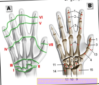
Outline of the hand
(Joint lines green)
I - Upper (proximal) wrist
II - Lower (distal) wrist
III - carpal metacarpal
Joints
IV - metacarpophalangeal joint
V - middle finger joint
(missing on thumb)
VI - interphalangeal joint
VII - thumb joint
- Distal phalanx -
Phalanx distalis - Phalanx -
Phalanx media - Phalanx -
phalanx proximalis - Metacarpal bones - Metacarpals
- Trapezoidal leg - Trapezium
- Trapezoid leg - Trapezoid bone
- Head leg - Os capitatum
- Hook leg - Hamate bone
- Scaphoid bone of the hand -
Scaphoid bone - Moonbone - Lunate bone
- Triangular leg - Os triquetrum
- Pea bone - Os pisiform
- Sesame bone - Os sesamoideum
- Cubit - Ulna
- Spoke - radius
You can find an overview of all Dr-Gumpert images at: medical illustrations
Illustration muscles of the hand
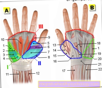
Hand muscles
I - muscles of the ball of the little finger
Hypothenary group (green)
II - muscles of the ball of the thumb
Thenar muscles (blue)
III - Metacarpal muscles (red)
- Little finger spreader -
Abductor digiti minimi muscle - Short little finger flexor -
M. flexor digiti minimi brevis - Short palm tendon tensioner -
Muscle palmaris brevis - Little finger counteracting device -
M. opponens digiti minimi - Thumb pull -
Muscle abductor pollicis - Short thumb flexor -
Muscle flexor pollicis brevis - Short thumb spreader -
Muscle abductor pollicis brevis - Thumb counter -
Muscle opponens pollicis - Spinal muscles -
Lumbrical muscles - Palm side intermediate
bone muscles -
Palmar interossei muscles - Elbow-sided hand flexor -
Flexor carpi ulnaris muscle - Spoke-sided hand flexor -
Flexor carpi radialis muscle - First back intermediate
bone muscle -
Muscle interosseus dorsalis I - Long thumb stretcher -
Extensor pollicis longus muscle - Short thumb straightener -
Extensor pollicis brevis muscle - Short spoke-side hand straightener -
Extensor carpi radialis brevis muscle - Long spoke-side hand straightener -
Extensor carpi radialis longus muscle - Backhand intermediate
bone muscles -
Dorsal interossei muscles - Finger extensor -
Extensor digitorum muscle - Klenfingerstrecker -
Extensor digiti minimi muscle - Extensor tendon strap -
Retinaculum musculorum extensorum - Ellenside hand extensor -
Extensor carpi ulnaris muscle
You can find an overview of all Dr-Gumpert images at: medical illustrations
Figure carpal bones
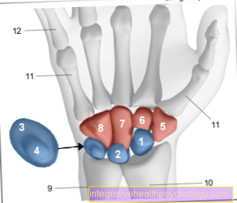
The eight carpal bones
Upper (proximal) row - blue
Lower (distal) row - red
- Scaphoid bone of the hand -
Scaphoid bone - Moonbone - Lunate bone
- Triangular leg - Os triquetrum
- Pea bone - Os pisiform
- Large polygonal leg
(Trapezoidal leg) - Trapezium - Small polygonal bone
(Trapezoidal leg) -
Trapezoid bone - Head leg - Os capitatum
- Hook leg - Hamate bone
- Cubit - Ulna
- Spoke - radius
- Metacarpal bones - Metacarpals
- Phalanx - Ph. Proximalis
You can find an overview of all Dr-Gumpert images at: medical illustrations
function
The wrist is a so-called Ellipsoid joint (Egg joint with two main axes, similar to a ball joint).
All partial joints of the wrist function as one unity and enable the complex movements of the wrist. They enable one diffraction (Flexion) towards the palm of the hand (Palmar flexion) by approx. 80 °, as well as the Elongation (Extension) Towards the back of the hand (Dorsiflexion) by about 70 °.
In addition, the wrist allows Splaying movements (Abduction) Towards the thumb (Radial abduction) and towards the little finger (Ulnar abduction) by approx. 30 to 40 °.
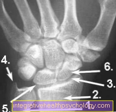
X-ray wrist
- Cubit (Ulna)
- Spoke (radius)
- wrist
- Stylus process (Ulna styloid process)
- Moonbone (Lunate bone)
- Scaphoid (Navicular bone)
Proximal wrist
The proximal wrist, which is closer to the center of the body, is formed by the articular surfaces of:
- Cubit (Ulna)
- spoke (radius)
- body-hugging series of Carpal bones (proximal carpal bones) consisting of the 3 carpal bones navicular bone (Scaphoid bone), Moonbone (Lunate bone) and triangular leg (Os triquetrum).
Together they make that Radiocarpal articulation. The capsule the joint is slack and thin.
On the side of the back of the hand (dorsal) the joint capsule is through different Tapes reinforced. The collateral ligaments between the carpal bones and the ulna or radius (radius) the stability. In addition, 2 ligaments between the carpal bones and the spoke from above and below strengthen the wrist (Radiocarpal ligament palmar and dorsal).
The entire joint is elliptical built up (ellipsoid joint or egg joint) and thus allows 2 different movements (2 degrees of freedom): The diffraction (Palmar flexion) and elongation (Dorsiflexion), as well as the splay in two directions (Ulnar abduction and Radial abduction).
The proximal wrist is mainly used for flexion (Palmar flexion), while in the distal wrist especially the Elongation (Dorsiflexion) takes place.
Distal wrist

The one away from the middle of the body distal wrist is used by the two rows the carpal bone (proximal and distal row) educated.
This creates a picture between the individual carpal bones Joint space, which one S-shaped is constructed.
There is also one here Joint capsule. This is rather tight on the inside of the hand, but rather slack on the back of the hand.
The distal wrist adjusts due to its structure toothed hinge joint which by its curved course, the ligaments and joint capsule severely restricted in its movement is. Together with the proximal wrist, it acts as a functional unit.
While the proximal wrist is primarily involved in flexion (palmar flexion), the distal wrist is particularly involved in the Stretching (dorsiflexion) instead of.
Articulationes intercarpales
This is the individual small joints between the carpal bones of a number. The individual bones are different Ligaments fixed tightly (Ligg. Intercarpalia interossea). The connections of the distant (distal) Line (Amphiarthroses).
Articulationes carpometacarpales
The distant (distal) Row of carpal bones forms together with the body-near (proximal) Finger bones (Ossa metacarpi 2- 5) stiffened joints (Amphiarthroses). These joints are drawn from the side of the palm (palmar) and the side of the back of the hand (dorsal) by tight ligaments fixed (Ligg. Metacarpalia dorsalia, palmaria and interossea).
The thumb joint is an exception here and is therefore significantly more flexible than the other individual fingers.
Metacarpophalangeal joint
in the Thumb saddle joint (Articulatio carpometacarpalis pollicis) interact the large polygonal leg (Trapezium) and metacarpal (Os metacarpale 1) of the thumb.
Since it is a saddle joint, 3 different directions of movement of the thumb are possible (3 degrees of freedom):
- diffraction (Flexion) and Elongation (Extension),
- Splaying movements
- and the Facing the thumb against the other fingers (Opposition and reposition). This is especially necessary for the so-called tweezer grip and without the opposition movement a person would hardly be able to grasp.
Carpal band
Across the individual carpal bones that is tense Carpal band (Flexor retinaculum). It runs between the hook leg (Hamulus ossis hamatis) and the pea bone (Os pisiform) to the cusp of the large polygonal bone (Tuberculum ossis trapezi) and the navicular bone (Tuberculum ossis scaphoidei). This creates an imagination connective tissue canalthat as Carpal tunnel is referred to and the Median nerve leads in its midst.
Wrist disorders
This is particularly common in the wrist Carpal tunnel syndrome on. This is caused by pressure and overload on the median nerve, which is located in the tunnel of the carpal ligament (Retinaculum flexorum) runs. The reason for this pressure damage can be previous Wrist fractures, rheumatic diseases, or overuse be.
The most common injuries to the wrist are a Break in the part of the spoke near the wrist (distal radius fracture), as well as a Fracture of the scaphoid bone (Scaphoid bone). In principle, all wrist bones can break, but this is the most commonly affected.
Another common condition is one Tendinitis in the area of the tendon sheath compartments on the back of the hand. In addition, a Arthrosis in the metatarsophalangeal joint occur. This is then saved as a Rhizarthrosis designated.
The X-ray image, the Ultrasonic and the MRI of the hand used.
Wrist in pain

Wrist pain can have many different causes.
Often there is only one temporary pain in the wrist overload of the joint, especially after intense sporting activities that strain the wrist, repetitive movements, for example when gardening or a cramped position of the wrist over a long period of time, for example when working at the computer for a long time.
Pain in the wrist that can be traced back to one of the above causes usually goes away on its own after a short time if the wrist relaxed becomes. However, the symptoms can also have other causes.
If you fall, you often reflexively support yourself with your hands. This can be painful Upsetting or to one Broken bone come in the hand area. In addition, this one can also Torn ligament on the wrist cause.
Persistent pain in the wrist after a trauma should therefore be clarified by a doctor so that a serious injury can be recognized and treated at an early stage.
Often there is also an advancing one Wear and tear of the wrist cause of the pain. In the course of this, osteoarthritis develops in the wrist, which is accompanied by worsening pain and later also with one Restriction of movement connected is. Initially, patients with osteoarthritis of the wrist only feel pain when they put more strain on the wrist, later the pain also occurs when the patient is resting and becomes more and more severe over time. In this case, treatment is first carried out with pain relievers and anti-inflammatory drugs.
In the final stage of osteoarthritis, these measures usually no longer help, so that the joint either has to be replaced with a prosthesis or stiffened in order to achieve lasting freedom from pain.
People who work a lot with the computer are more likely to suffer from the so-called Mouse hand. In this case, the wrist hurts due to a cramped posture when using the mouse and keyboard. Special mousepads with a raised surface for the palm of your hand can help. Such overloading of the wrist can also lead to tendinitis, which can also cause pain in the wrist. Often the wrist is also swollen.
Another possible cause of wrist pain is that Carpal tunnel syndrome. This is a bottleneck syndrome in the area of the wrist, which leads to a narrowing of the median nerve under the connective tissue plate on the wrist. The increased pressure on the nerve leads to unpleasant tingling and numbness, as well as pain in the wrist, which occurs especially at night.
Tapping the inside of the wrist can also trigger electrifying pain (so-called Hoffmann-Tinel sign). Overall, there are many different causes for pain in the wrist, so that a doctor should be consulted if the symptoms persist.
You can find much more information on this topic at: Wrist pain
Sprained wrist
A wrist sprain occurs when it is severely overstretched, i.e. when the joint moves beyond the normal range of motion of the joint. As a result, ligaments and joint capsules are greatly stretched and, depending on the extent, can also tear.
A wrist sprain is very painful. The joint usually swells up a lot and a bruise can form if vessels were damaged when the joint was overstretched. If the pain is so severe that the joint can no longer be moved, it could also be a broken bone. Normally, if there is a sprain, the wrist can still be moved to a certain extent despite the severe pain.
The final diagnosis of the sprain is made by a doctor. If there is uncertainty as to whether a bone has not broken, an X-ray is usually taken for clarification.
If it is just a sprain, conservative therapeutic measures can be used. First and foremost, the wrist should be immobilized. All you need to do is wrap the joint with an elastic bandage or put on a wrist splint. In addition, the wrist should be cooled extensively and elevated. This will reduce swelling and pain. Cooling or decongestant ointments can also be used. As a rule, the symptoms should have subsided after two weeks. If it lasts longer or gets worse over time, there may be a more serious injury after all. In this case, a doctor should be consulted so that appropriate therapy can be initiated. This is the only way to avoid consequential damage to the wrist.
Athletes can do a lot to avoid a wrist sprain in the first place. It is important that the joints are sufficiently warmed up and stretched before strenuous exercise. As a result, you are better prepared for the coming stress and can better absorb the forces. Taping the wrists is also helpful, especially if a previous wrist injury has occurred. The bandage stabilizes the wrist and is less prone to injury.
You might also be interested in: Sprain on the thumb
wrist broken
Colloquially it is called a broken wrist when there is a fracture at the bottom of the wrist spoke (Radius) is present. This is one of the most common broken bones and affects all ages.
In old age, women are particularly affected because they are through osteoporotic changes are more prone to fracture. Overall, the most common cause of a broken spoke is direct violence, for example a fall on the outstretched hand, an impact trauma or a sports injury.
The break occurs immediately severe pain in the wrist area, which is accompanied by severe swelling and often also bruising. Sometimes the fracture is already visible on the outside when the ends of the bones stand apart and cause a deformity in the forearm. Otherwise, the diagnosis of the fracture is made using a X-ray image posed. The fracture gap is then visible.
In addition, the doctor can use the X-ray image to decide which therapy is appropriate for the type of fracture present. If the fracture ends of the bone are not displaced against each other, then it is often enough Forearm castused to fix the wrist. This is to prevent the fracture from slipping and the misaligned bone ends from healing together. If the break is postponed from the start, the pieces of bone must be brought back into their correct position. This is usually a operational approach necessary in which the bone fragments through Screws, Wires or plates be fixed again in their correct anatomical position. The bone can then heal. After several weeks to months (depending on the type of operation), the pieces of metal are usually removed again. As a rule, the plaster of paris remains in place on adults for between three and six weeks.
During follow-up treatment, it is important that physiotherapy exercises in order to restore the normal functioning of the wrist. Nevertheless, under certain circumstances, slight restrictions in the function of the joint may remain. However, these are usually not severe, so that no significant restriction in professional or everyday life is to be expected. Long-term complications from a spoke fracture are primarily the development of osteoarthritis or the occurrence of one Stunted growth with children.
Tap your wrist

To tape the wrist you need Kinesiotape strips of 3.75cm Width. The ends of the strips should be removed before gluing rounded as they won't come off as quickly that way.
To apply the tape, the affected arm is loosely placed on a table. It is easiest if a second person attaches the tape strips.
First, make a strip just in front of your wrist circular to the forearm wrapped. A second strip is around in the same way Palm and back of Hand glued just before the insertion of the fingers. These two strips are then connected to one another by the strips that are glued on below. First the back of the hand is glued.
It starts with a strip on the side of the thumb, which must also be the Thumb saddle joint should include. The other strips are attached next to it at regular intervals and each end at the circular strip in front of the wrist.
Then two strips of tape diagonal glued. The first starts on back of Hand at the level of the little finger, then runs across the back of the hand and ends at the circular strip of tape on the wrist on the side of the thumb. The second strip runs so that it crosses the first. It starts on the back of the hand on the thumb side and runs across the direction of the circular tape on the wrist on the little finger side.
If all these tape strips are stuck on the back of the hand, the same procedure is used with the Palm of the hand proceed. Finally, the hand and wrist are completely covered with horizontally glued tape strips so that the previously glued strips are no longer visible. The wrist is then sufficiently stabilized. Overall, when taping, care should be taken that the strips are not stretched too much, and ultimately the tape not too tight sits.
It shouldn't be found uncomfortable.
Summary
The wrist is a very complex jointwhich is made up of various partial joints composed. It consists of:
- a proximal joint close to the body
- and a distal wrist distal from the body.
- In addition, the individual carpal bones and their joint surfaces belong to the wrist in a broader sense.
Only in the cooperation of the individual partial joints is the great mobility of our wrist.
The articular surfaces are through numerous tapes fixed so that some joints have significantly less mobility (Amphiarthroses) than others.
Due to the complex structure of the wrist, the numerous structures and bone can the wrist hurt quickly will and is with it endangered with every fall on the hand. Because of the numerous activities that humans perform with the wrist every day, it is robust, but can also overexerted become and then to painful inflammation or wear and tear (arthrosis) to lead.
Illustration wrist pain
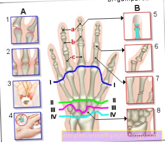
A - Chronic causes
B - Acute causes
- Rheumatoid Arthritis (RA) -
inflammatory disease
the joints - Arthrosis -
Joint wear - Carpal Tunnel Syndrome (KTS) -
constriction of the median nerve - Ganglion (upper leg) -
benign tumor formation - Torn ligament -
Joint ligament rupture - Finger dislocation -
Dislocated finger - Broken finger (finger fracture) -
a - distal
b - medial
c - promaximal - Wrist hernias
(here scaphoid fracture)
I - I - metatarsophalangeal joint -
Articulatio metacarpophalangea
II - II - Carpal-metacarpal joints -
Articulationes carpometacarpales
III - III - Lower wrist -
(distal)
Articulatio mediocarpalis
IV - IV - Upper wrist -
(proximal)
Radiocarpal articulation
You can find an overview of all Dr-Gumpert images at: medical illustrations