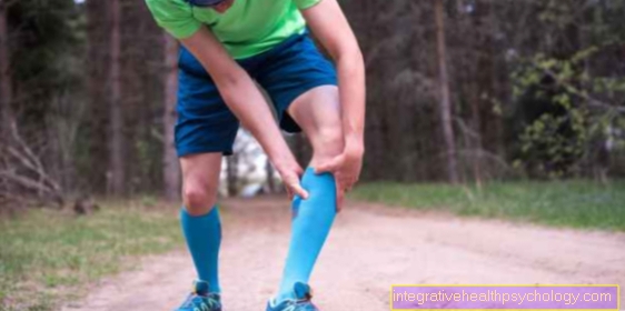
Cartilage damage in the knee
causes
Cartilage damage in the knee is quite common. Most of the time, the damage is caused by wear and tear. On the one hand, this wear and tear occurs as part of a completely natural aging process. The expert describes the result as osteoarthritis (chronic degenerative joint disease).
You might also be interested in this topic: Cruciate ligament overstretched
The knee joint has to carry almost our entire body weight and is subjected to many other loads and movements every day.It is therefore not surprising that the main risk factors for cartilage damage in the knee are overweight and incorrect or excessive loads on the knee joint, such as certain types of sport, and of course advanced age. From the age of 70, almost everyone has more or less pronounced osteoarthritis of the knee joint.
In addition, misalignments in the knee, such as knock knees or bow legs, can cause increased wear and tear on the cartilage. Injuries such as tears of the inner ligament or cruciate ligament are less common at the beginning of a process that ultimately leads to damage to the cartilage in the knee.
Bald cartilage describes the condition when there is no more cartilage. For more information, read the article at: Bald cartilage - is it dangerous?
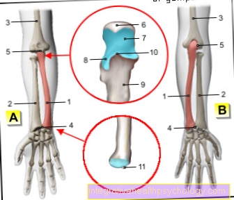
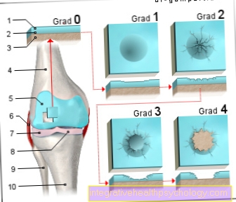
Figure cartilage damage in the knee joint

- Articular cartilage
(hyaline cartilage) -
Cartilago articularis - Cartilage remodeling zone
in bones -
Zona ossificationis - Joint body (articular gnar
of the femur) -
Femoral condyle - Femur -
Femur - Articular cartilage -
Cartilago articularis - Outer band -
Ligamentum collaterale fibulare - Outer meniscus -
Lateral meniscus - Inner meniscus -
Meniscus medialis - Fibula - Fibula
- Shin - Tibia
You can find an overview of all Dr-Gumpert images at: medical illustrations
diagnosis
The diagnosis arthrosis offers the doctor very classic changes, especially in the X-ray image become clearly visible. The joint space is narrowed because the cartilage is worn out. As a reaction to this, the surrounding bone has hardened and new bone parts are sometimes formed in order to enlarge the contact surface and thus start an attempt to absorb the increased pressure caused by the missing cartilage in this way.
To the cartilage damage to the knee joint or a Cartilage damage behind the kneecap To be able to classify better, one uses the classification according to Outerbridge, in which grades 0 to 4 are differentiated. (see below)
- Grade 0: no existing cartilage damage;
- Grade 1: the cartilage is completely preserved, but softens under pressure;
- Grade 2: the cartilage is superficially a little separated;
- Grade 3: the cartilage is torn down to the bone;
- Grade 4: the cartilage is completely lost down to the bone, so the bone is exposed.
However, the extent of the patient's complaints does not always have to be directly related to the extent of the changes in the joint, which is why it is always important not to rely on the imaging alone, but to take an extensive anamnesis with the patient.
However, cartilage damage to the knee cannot be adequately assessed using the X-ray image. The best method of examining cartilage damage is this MRI of the knee.
The MRI of the knee is an examination method, which in addition to the cartilage damage also damage to the meniscus (Inner meniscus and outer meniscus), such as Damage to the posterior and anterior cruciate ligament can represent temporally.
You can find further information under our topic: MRI knee

I would be happy to advise you!
Who am I?
My name is dr. Nicolas Gumpert. I am a specialist in orthopedics and the founder of .
Various television programs and print media report regularly about my work. On HR television you can see me every 6 weeks live on "Hallo Hessen".
But now enough is indicated ;-)
The knee joint is one of the joints with the greatest stress.
Therefore, the treatment of the knee joint (e.g. meniscus tear, cartilage damage, cruciate ligament damage, runner's knee, etc.) requires a lot of experience.
I treat a wide variety of knee diseases in a conservative way.
The aim of any treatment is treatment without surgery.
Which therapy achieves the best results in the long term can only be determined after looking at all of the information (Examination, X-ray, ultrasound, MRI, etc.) be assessed.
You can find me in:
- Lumedis - your orthopedic surgeon
Kaiserstrasse 14
60311 Frankfurt am Main
Directly to the online appointment arrangement
Unfortunately, it is currently only possible to make an appointment with private health insurers. I hope for your understanding!
Further information about myself can be found at Dr. Nicolas Gumpert
Classification according to severity
Cartilage damage in knee grade 1
Of the Grade 1 cartilage damage according to the classification according to Outerbridge corresponds to a slight damage to the knee joint.
Generally these Cartilage damage in technical language too Chondropathies called. In order to classify cartilage damage as first degree, a lesion does not necessarily have to be recognizable.
The surface of the cartilage is still intact here and only slight softening and discoloration of the Cartilage tissue or detect small superficial cracks and fissures. Large fraying or tears are not visible.
The exact degree of cartilage damage is determined by athroscopy. In the case of the first-degree damage, the cartilage appears softened and more easily injured. Of the Grade 1 cartilage damage arises from overstressing the knee joint and often causes only mild symptoms and rarely pain, so that surgical therapy is usually not sought.
Cartilage damage in knee grade 2
The cartilage of the knee joint consists of different layers that differ slightly in their composition and ensure the stability and resilience of the cartilage.
At a Grade 2 cartilage damage to Outerbridge there are significantly deeper tears and fraying compared to grade 1.
In some parts of the cartilage, such a tear can reach so deep into the cartilage that half of the tissue is affected. However, the bone of the thigh and tibia, which lies under the cartilage, is not affected by the damage and is still covered by cartilage.
From a therapeutic point of view, it is very important to distinguish between a simple and deep tear and a large-scale rubbing of the cartilage. The damage mentioned first is usually not changed even with imaging follow-up examinations of the cartilage damage and therefore does not necessarily require surgical therapy.
On the other hand, abrasion of the cartilage is unfortunately often characterized by further progressive damage, which leads to an increase in symptoms.
In this case, the cartilage cannot recover from the damage on its own and so the cartilage damage usually quickly develops into grade 3 cartilage damage.
Cartilage damage in knee grade 3
The damage to the Third degree knee cartilage characterizes a lesion that affects over half the layer thickness.
This can be a deep tear that occurs either as a result of permanent overuse of the knee joint or often as a result of a traumatic event.
If the deep cartilage damage is more of a broad abrasion, this already indicates that the bone that is located below the cartilage and is involved in the formation of the knee joint will soon be exposed. Depending on the cartilage layer affected, the third-degree damage can be divided into three subclasses according to IRCS.
- In the first class the defect does not extend to the calcifying layer of the cartilage
- in the second grade this layer is also affected by the lesion and in the third class the defect even extends into the subchondral layer, which represents the boundary between cartilage and bone.
- Of the third-degree cartilage damage In any case, it is an indication for surgical treatment. There are various surgical techniques to repair the defect and to relieve the patient from pain.
Cartilage damage in knee grade 4
The Grade 4 cartilage damage corresponds to the greatest damage to the knee joint cartilage Outerbridge.
Not only does the lesion affect the completely damaged cartilage itself, but it has spread to the neighboring structure, in this case to the bone involved in the formation of the knee joint.
In this context, one speaks of damage in the form of Ulcers, since these are defects that affect all layers of a tissue and, starting from the top layer, spread out into the depths.
There is maximum joint wear and the bone itself can also be deformed and show signs of wear.
This can for example Ground grooves due to the constant mechanical stress in the form of friction in the knee joint. The consequences of the fourth-degree cartilage damage are severe pain in the affected patient and significantly reduced resilience.
This severe cartilage damage should be treated urgently in order to avoid further damage to the bone.
Symptoms
Those affected usually complain primarily about pain. In the beginning, these are usually only found under stress, but also increasingly under rest conditions over the course of the day. At an advanced stage, the knee joint can suffer from functional impairments, which are sometimes accompanied by an effusion of the knee joint (in its chronic form also called Baker's cyst) or deformations of the joint.
Find out more about: Acute knee pain - that may be behind it
Symptom of swelling and water in the knee
A swelling of the knee joint manifests itself as a Increase in size of the joint and can be recognized very easily by comparing it with the healthy side.
This swelling can be due to the retention of fluids such as water, pus or blood act. In the event of water retention, the water present in the knee also has a variety of possible causes and is very uncomfortable for the patient.
The accumulation of water occurs due to mechanical or inflammatory Damage to the structures involved in the knee joint and does not occur in a healthy knee joint.
In addition to the cartilage and bone damage, it can also originate from the muscles, ligaments and vision or from the bursa of the knee joint.
In order to establish a diagnosis, it is important to distinguish whether the swelling was preceded by trauma and the period in which it developed.
In the case of water accumulation in the knee joint, one is suitable for researching the cause and for the purposes of the first symptomatic treatment Puncture same.
This can reduce the pressure in the joint space, any blood or pus admixture is recognized and the puncture can then be searched for various pathogens and bacteria.
The cause of the knee swelling should be treated as further therapy. In addition to surgical measures, drug-based pain therapy, exercise therapy as part of a physiotherapeutic treatment and strengthening of the leg muscles, which is particularly recommended for slight cartilage damage, can be considered or prescribed.
Appointment with a knee specialist?
I would be happy to advise you!
Who am I?
My name is dr. Nicolas Gumpert. I am a specialist in orthopedics and the founder of .
Various television programs and print media report regularly about my work. On HR television you can see me every 6 weeks live on "Hallo Hessen".
But now enough is indicated ;-)
The knee joint is one of the joints with the greatest stress.
Therefore, the treatment of the knee joint (e.g. meniscus tear, cartilage damage, cruciate ligament damage, runner's knee, etc.) requires a lot of experience.
I treat a wide variety of knee diseases in a conservative way.
The aim of any treatment is treatment without surgery.
Which therapy achieves the best results in the long term can only be determined after looking at all of the information (Examination, X-ray, ultrasound, MRI, etc.) be assessed.
You can find me in:
- Lumedis - your orthopedic surgeon
Kaiserstrasse 14
60311 Frankfurt am Main
Directly to the online appointment arrangement
Unfortunately, it is currently only possible to make an appointment with private health insurers. I hope for your understanding!
Further information about myself can be found at Dr. Nicolas Gumpert
therapy
The problem with cartilage damage is that the human body is only able to a very limited extent to Cartilage tissue to regenerate again. This is because this type of tissue is not supplied by nerve cells and blood vessels, which are, however, of great importance for the healing process. One assumes that only about 4% of the cartilage cells can be renewed, which, however, sometimes dated Age depends.
Most of the time, the damage will tend to increase rather than improve over time. For this reason, the aim of treatment is rather to prevent further damage from occurring. First of all, it is important that everyone Risk factors be switched off or reduced as far as possible. Obesity should be reduced, stressful sports and other Overexertion should be avoided Underlying diseases or Misalignments should be eliminated. (see also: Knee osteoarthritis and exercise)
In addition, depending on the x-ray findings and the level of suffering of the patient, one decides which therapy is most useful.
Of great importance is that physical therapy, a conservative (non-operative) therapy. In the case of less pronounced changes, this can be sufficient to enable an affected person to lead a symptom-free life. The is particularly popular Arthroscopy (Arthroscopy). During this procedure, bone or cartilage parts can be removed, the cartilage smoothed and the joint cleaned and flushed (Lavage or. Debridement). Can accompany Painkiller and or Shoe insoles are used.
If these measures are not sufficient to significantly improve the symptoms, there are still some newer methods of dealing with cartilage damage in the knee: Especially with younger patients, it is advisable to place healthy cartilage tissue in the knee transplant. There are also brand new drugs on the market that specifically inhibit certain inflammatory triggers that are responsible for damage to the knee cartilage. Although these are currently still being tested and are very expensive, they promise great success.
Finally, it should be mentioned that cartilage cultivation is currently being researched. So you are now able to grow cartilage cells from blood stem cells in the laboratory and such Cartilage cultures and subsequent Transplants are already being carried out successfully in some areas of Germany.
Healing of the cartilage damage in the knee joint
Depending on size, localization and depth the cartilage damage and depending on the patient's complaints, various surgical therapies are carried out, the aim of which is to use the body's self-healing tendency in order to achieve the best possible stabilization of the cartilage tissue and to enable the patient to be free of symptoms.
However, healing and regeneration of the original cartilage tissue is not possible. Rather, it can be done in different ways Substitute substances be used for the lost cartilage.
The rate of self-healing is highest in young people who have suffered a cartilage injury from a traumatic event.
In addition to drug therapy and exercise, no surgery is required here. By the process of the so-called Microfracturing For small cartilage defects, a hole can be drilled in the bone, which is located below the cartilage.
This leads to a bleeding into the cartilage and an accumulation of stem cells and donor factors, which cannot restore the cartilage and thus do not achieve complete healing, but these cells stimulate the formation of Fiber cartilage and Scar tissue at the defective point, which in the best case leads to a relief of the patient's symptoms.
In addition to this surgical technique, you can also the body's own cartilage (OATS) or only cartilage cells can be transplanted into the knee joint (ACT) to replace the defective cartilage.
Scraping and smoothing of the surface is also possible in the case of slight lesions, but this reduces the thickness of the entire cartilage.