MRI for a stroke
What is an MRI for a stroke?
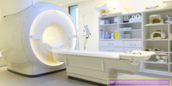
MRI (magnetic resonance imaging) is a diagnostic procedure that uses magnetic fields and radio waves to create an image.
With the MRI, even small strokes can be displayed very well and, above all, much earlier than with the CT (computed tomography). In this article, you will find out why the CT examination is still the most important examination when a stroke is suspected.
As with any procedure, there are also disadvantages in this case, for example the high costs or the more difficult monitoring options for seriously ill patients.
Get basic information on:
- Stroke.
- This is how a stroke is diagnosed
Why do you do an MRI if you have a stroke?
The most important imaging requirement in general for acute ischemic (decreased blood flow) stroke is to exclude cerebral haemorrhage.
Furthermore, the images serve to evaluate the degree and spatial extent of the stroke and thus the brain damage. This enables the chances of success of the therapy to be assessed.
Imaging is also important to rule out stroke mimics (other causes that cause stroke-like symptoms). These include e.g.
- subacute encephalitis
- Seizures
- a space claim
- an acutely symptomatic CSF circulatory disorder
When do you do an MRI if you have a stroke?
There are several indications for an MRI.
In an acute situation, it is used when the time window is unclear. This is especially the case with wake-up strokes, when symptoms are noticed when you wake up and the exact time at which symptoms began remains unclear. Even if the onset of symptoms is> 4.5 hours, MRI is used as the basis for revascularizing therapy (improvement of the blood flow to less well supplied tissues).
In the process, the MRI is used to rule out other possible diagnoses (differential diagnoses), so-called stroke mimics. In addition, the infarct pattern can be displayed very well in this way.
The MRI has additional benefits in the following aspects:
- Early visualization of lesions
- Assessment of the tissue at risk (penumbra: this refers to brain tissue that is functionally disturbed by a lack of oxygen in the event of a stroke, but can be saved from cell death by revascularizing therapy.)
- High sensitivity even with small heart attacks
Can you detect every stroke with an MRI?
MRI has great resolving power so that you can detect even small strokes. However, there are tiny strokes and bleeding that the MRI escapes.
If MRI is used as an imaging method in the acute phase of ischemic stroke, it should be noted that no diffusion disorder (disruption of substance transport) can be measured in the MRI in around 20 to 35% of patients. This applies to so-called clinically manifest ischemia and non-transitory ischemic attacks.
In this case, the clinical findings, not the imaging results, are decisive. A lack of detection by MRI is no reason to doubt the well-founded clinical diagnosis of a stroke. There is also no reason to change the guideline-based therapy. This does not affect the outcome of these patients.
MRI or CT of the head - which is better?
There is no general answer to this question. Each process has its advantages and disadvantages.
MRI is superior to CT in detecting ischemia (decreased blood flow). It can prove this earlier and even at a smaller size. In addition, it shows ischemia of certain areas of the brain with greater certainty than CT of the brain stem. The MRI makes better statements about the development / cause of infarcts and shows a clear superiority in the detection of stroke mimics (other causes that cause stroke-like symptoms).
Disadvantages of MRI are, for example, the long duration of the examination, the high costs, the more difficult monitoring options for critically ill patients and the delays in the diagnosis. This includes, for example, patients with cardiac pacemakers or other metallic implants, even if these days this is usually not a contraindication to an MRI scan.
Despite the considerable number of advantages of MRI mentioned above, CT is considered to be the most important examination in the diagnosis of a stroke. In accordance with guidelines, a native CT should be used to rule out intracranial (in the skull) bleeding. From a purely clinical point of view, it cannot be reliably differentiated from an ischemic stroke. However, intracranial hemorrhage is a contraindication to initiating intravenous lysis therapy such as that used in ischemic stroke.
The decisive advantage of CT compared to MRI is the significantly lower effort and the shorter duration of the examination, which is particularly important in an emergency situation. In addition, the radiation dose with the new computer tomographs has become so low that the radiation exposure, at least in emergency diagnostics, is no longer an argument against CT.
Learn more about the Difference between the MRI and CT.
Do you need contrast media?
Contrast media are used to better represent structures. The MRI images are black and white and since there are not an infinite number of shades of gray, it is sometimes difficult to differentiate between different tissues.
Diffusion MRI is particularly important for diagnosing a stroke. Here, areas are shown in which diffusion (material transport) is restricted and the cells are thus irreversibly damaged. No contrast media is required for this examination.
In each individual case, the radiologist decides whether or not to use contrast media.
Read whether a MRI with contrast agent is dangerous.
When do you do a follow-up?
A follow-up based on imaging is rather unusual.
Regular aftercare after a stroke begins with inpatient or outpatient rehab, depending on the severity of the stroke. The aim is to restore functions such as language and motor skills through physiotherapy, speech therapy and occupational therapy. The patient trains to master everyday (professional) life independently again.
Learn more about the Healing after a stroke.
Does an MRI help better assess the risk of stroke?
An MRI is not used prophylactically to determine the risk of stroke. It is only used if there is justified clinical suspicion.
It is more effective to minimize the known risk factors. Which includes:
- Restriction of smoking
- Adjustment of blood pressure
- Treatment of irregular heartbeat
- Change of diet
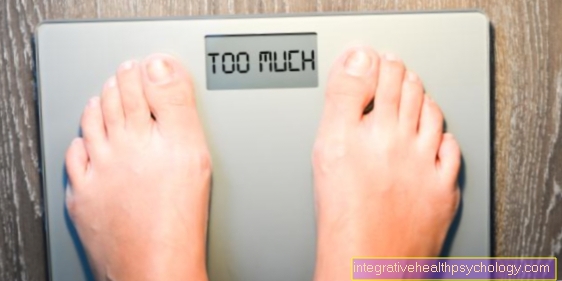
- Loss of excess weight
- light sporting activity
- Avoiding stress
Read how to do one Prevent stroke can.