Pubic bone
General
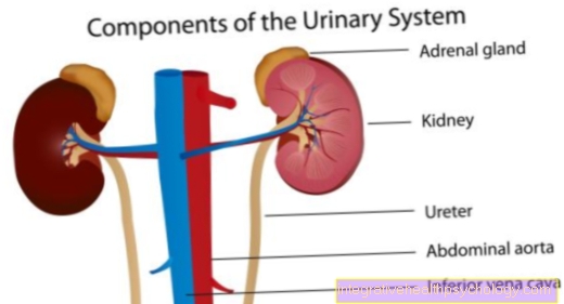
The pubic bone (lat. Pubis) is a flat bone and part of the pelvis. It occurs on both sides of the pelvis and is connected in the midline by the pubic symphysis. It is placed in a pubic body (Corpus ossis pubis) and two pubic branches each (Ramus superior and inferior of the pubic bone). The pubic bone is part of the hip joint and the origin of some muscles that help build the pelvic floor.
construction
The pubic bone is an angular, flat bone that is part of the pelvis. It is present on both sides of the pelvis. The pubic bones are connected to one another via a fiber cartilaginous connection, the pubic symphysis. Since this connection is a fiber cartilage, the two pubic bones can easily be moved against each other. These shares are called Corpus ossis pubis designated.
This is followed by two pubic branches, the Ramus superior (upper branch) and the Ramus inferior (lower branch) of the pubic bone. These represent the connections to the adjacent bones and are part of the hip joint.
To the front and up is the upper pubic branch (Ramus superior ossis pubis) with the iliac bone (Os ilium) connected, the lower pubic branch (Ramus inferior ossis pubis) with the ischium (Os ischii). These connections are bony and therefore cannot move against each other.
The pubic bone forms the anterior portion of the Acetabulum (Acetabulum). This in turn forms together with the Femoral head (Head femoris) the hip joint. The anterior border of the pubic bone is called the Pubic crest (Pecten ossis pubis) designated. The midline, i.e. the connection between the two pubic bones, is called the Symphysis designated.
The pubic crest bears a small hump at its transition to the midline, which as Pubic tuberosity referred to as. It serves as the starting point of the Inguinal ligament. At the lateral edge of the pubic ridge this ends in another hump, the Eminentia iliopubica. This represents the border with the iliac bone.
In addition, the pubic bone limits the anterior arch of the Obturator foramen. This is supported by the Obturator externus muscle and internus locked. This leaves only a small channel, the Obturator canal. The Obturator nerve, as well as the Vena and Obturator artery.
Musculature
It arises from the pubic bone Pubococcygeus muscle and the Puborectalis muscle. Both are parts of the Levator ani muscle and thereby form the Pelvic floor muscles.
Furthermore, the Transversus perinei profundus muscle on the pubic bone. This is part of the Urogenital diaphragm, which in turn is involved in the structure of the pelvic floor.
Summary
The Pubic bone (Pubis) is part of the bony Basin and is available once on each side of the pelvis. It's part of the Hip joint and forms with the Iliac bone (Os ilium) and the Ischium (Os ischii) the Acetabulum. The two parts are connected by a fiber cartilage in the middle (Symphysis) and thus can be moved against each other. Furthermore, the pubic bone is the origin of some muscles, which are parts of the Pelvic floor form.
.jpg)