molar tooth
General
The molars are mainly used to grind the food that has been pre-ground by the incisors. The molars are divided into two groups:
- anterior molars (Dentes premolars, premolars) and the
- posterior molars (Dentes molares)
The front molars (premolars)
The anterior molar / the Premolars are also called Vormahlzahn or Bicuspid (from Latin to "twice" and cuspis "Tip").
In contrast to the posterior molars, the premolars also have milk tooth predecessors, which are before the Change of teeth serve to grind the food.
Today people only have 2 premolars (front molars) per half of the jaw, these are with the tooth formula as 14, 24, 15, 25, 34, 44, and 35, 45 designated. Our mammalian ancestors originally had twice as many premolars, i.e. four front molars per half of the jaw.
The individual premolars have two to three cusps in humans Dental crownmaking the function of milling possible.
The lower premolars show a very pronounced crown alignment. The number of each Tooth roots and root canals varied between each premolar. Teeth 14 and 24 usually have two tooth roots and two tooth canals and two tooth cusps on their surface.
Teeth 15 and 25, however, have only one tooth root and one or two tooth canals. These too have two humps on their surface. Teeth 34 and 44 have a root and a root canal, rarely two root canals. They also have two humps. Teeth 35 and 45 have only one root and one root canal, but two or three tooth cusps.
Deviations from this scheme are possible.
The rear molars
The back molars belong to the large molars and are in the childlike teeth not to be found. For this reason, they are popularly known as incremental teeth.
They are also known as molars and are particularly large and powerful. They wear pronounced Humps (Tubercle) and dimple (Fissures).
With humans belong the 6., 7. and 8th tooth to the molars, which means that humans have three large molars per half of the jaw, making a total of 12 posterior molars.
The first posterior molar (6th tooth) usually erupts at the age of 6 and is therefore known as the six-year-old molar. The second rear molar (7th tooth) does not appear until the age of 12, the last molar (8th tooth) only breaks through in adulthood between the ages of 18 and 25.
For this reason it is also called Wisdom tooth designated. As with the premolars, the number of tooth roots and root canals as well as the cusps between the individual molars varies. The two teeth 16 and 26 have three tooth roots, four root canals and four tooth cusps.
Teeth 17 and 27 each have three tooth roots and root canals as well as four tooth cusps. Teeth 36 and 46 have two tooth roots and three root canals, but five tooth cusps. The teeth 37 and 47 are constructed in the same way, but only have four tooth cusps.
Teeth 18, 28 and 38, 48 do not have a fixed number of roots, canals and cusps and are very different from one individual to the other. Even with the large rear molars, deviations from this scheme are possible.
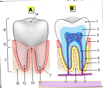
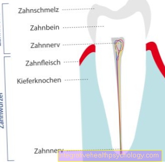
a - tooth crown - Corona dentis
b - tooth neck - Cervix dentis
c - tooth root - Radix dentis
- Tooth enamel -
Enamelum - Dentin (= dentine) -
Dentinum - Tooth pulp in the tooth cavity -
Pulp dentis in Cavitas dentis - Gums -
Gingiva - Root canal
- Cement -
Cementum - Root skin - Periodontium
- Opening of the tooth root tip -
Foramen apicale dentis - Nerve fibers
- Alveolar bone (tooth-bearing
Part of the jawbone) -
Pars alveolaris
(Alveolar process) - Blood vessels
- Tooth root tip -
Apex denitis - Point of division of the tooth roots
(Fork) - Bifurcation - Tooth furrow
You can find an overview of all Dr-Gumpert images at: medical illustrations
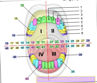
I - right upper jaw -
1st quadrant (11-18)
II - left upper jaw -
2nd quadrant (21-28)
III - lower jaw left -
3rd quadrant (31-38)
IV - lower jaw right -
4th quadrant (41-48)
- 1. Incisor -
Dens incisivus I - 2nd incisor -
Dens incisivus II - Canine tooth -
Dens caninus - 1st molar tooth
Anterior tooth (premolar) -
Dens premoralis I. - 2. Anterior molar
Anterior tooth (premolar) -
Dens premoralis II - 1st molar tooth -
Dens molaris I - 2nd molar tooth -
Dens molaris II - Wisdom tooth (= 3rd molar) -
Dens molaris tertius
(Dens serotinus)
1st - 3rd are front teeth
(3 per quadrant)
4th - 8th are molars
(5 per quadrant)
You can find an overview of all Dr-Gumpert images at: medical illustrations
Pulling the molar
Under the Pulling a tooth or the molar one understands that Extract of the entire Tooth or. Molar with connected Gums and Bone material.
A anesthesia is usually not necessary. If there is a need there is one local anesthesia possible. The injection as such can also be painful. First, the molar is loosened by the dentist using a lever. Then the affected tooth is carefully removed with a forceps instrument jaw brought out. Usually you feel a kind of pressure in the jaw. There should be no pain.
Important after the actual pulling of the tooth is the cleaning or. disinfection.
On a thorough oral hygiene should always be respected. If you have a tendency to bleed, a hemostatic tamponade inserted, which after approx. a week can be removed. After this time, that too Sutures removed if the affected oral mucosa has to be sutured.
The removal of the molar is often necessary for orthodontic reasonsto create the space needed for it Deformities can be avoided. Extracting a molar is a routine procedure for the dentist.Nevertheless, caution is advised when pulling, as adjacent teeth should not be damaged. In rare cases, even a Hairline crack in the jawbone arise. In this case, complaints do not become noticeable until a few weeks later.
Broken tooth
The Abort or canceling part of the molar tooth is not uncommon and often results from the enormous physical strainthat this tooth is exposed to. It is advisable to see a dentist as soon as possible. If the molar or part of it breaks off, this also means that the The root of the tooth is still in the jaw.
Well surrender 2 optionswhich the dentist has to weigh. On the one hand, a cut in Gums the Root to be taken out and a Implant or a prosthesis can be used. On the other hand, if possible, the Keep the root and a Crown to be put on.
Should a possible after clarification Tooth restoration be carried out, this first implies a thorough one Root canal treatment ahead. The decisive factor for the type and extent of the molar restoration is Length of the broken tooth part. If the Enamel that's often enough Grinding the tooth with a subsequent Glaze with the help of a varnish. In the case of a deeper tooth breakage, restoration is usually carried out using a Inlays.
Toothache

Toothache in molars is extremely uncomfortable pain. They have a massive impact on the patient's everyday life. Normal chewing of food is hardly possible anymore, which is why a clarification by the dentist is necessary in order to find out the cause of the pain.
The most famous trigger for toothache is tooth decay. These occur when caries forms a hole in the tooth that attacks the nerve tip. The nerve marrow of the tooth (pulp) withdraws further and becomes deserted.
If it turns out that the molar was not preserved, the dentist will pull the molar. However, this should be seen as the last option, as the molar is extremely important for the process of chewing.
Rather, one tries to eliminate the defect first. If this sits under the crown, an attempt is then made to put it back on again or even to have a new crown made (after an impression). An X-ray is also part of the dentist's clarification of the cause. If tooth decay was discovered or treated too late as the cause, one will occur Pulpitis. There is inflammation of the nerve marrow here. The resulting swelling with the accompanying pressure leads to permanent, pulsating pain in the area of the molar. In rare cases, these diseases can also result from injuries to the jaw. Other causes would be abscesses, periodontitis or the falling out or loosening of dentures, which means that the ground tooth is now sensitive to food or liquids.
Read more on the topic: Toothache
Formation of the molars in babies
In the development of the Teeth in young children pain often occurs. It is important to know that Incisors cause little to no pain when breaking through the gums. Essential The growing or the formation of the molars in small children is more painfulbecause they have a large and blunt surface. The molars usually make up between the 12th and 15th month of life on the way from jaw through the still soft gums. This process can up to the 18th month of life stop. If the molar comes through the gums, it is more the pressure that causes the pain. Also a Swelling of the cheeks is to be observed.
A common side effect is fever. This is because Germs causing fever (Pyrogens) now get more easily into the organismbecause it is precisely at this time that children hands in the mouth to take. It applies above all Keep calm and the child a lot during this time To pay attention. As a rule, the molars can be fully seen after they are 24 months old.