Function of the cerebellum
Synonyms
Medical: Cerebellum (lat.)
English: cerebrellum
introduction
The fact that the cerebellum contains nerve cells in particular, which have an inhibiting effect, gives an idea of its function. The cerebellum serves - to put it briefly at the beginning - to control movement sequences, primarily to limit movements so that they are regulated and do not become excessive.

Illustration brain
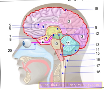
Cerebrum (1st - 6th) = endbrain -
Telencephalon (Cerembrum)
- Frontal lobe - Frontal lobe
- Parietal lobe - Parietal lobe
- Occipital lobe -
Occipital lobe - Temporal lobe -
Temporal lobe - Bar - Corpus callosum
- Lateral ventricle -
Lateral ventricle - Midbrain - Mesencephalon
Diencephalon (8th and 9th) -
Diencephalon - Pituitary gland - Hypophysis
- Third ventricle -
Ventriculus tertius - Bridge - Pons
- Cerebellum - Cerebellum
- Midbrain aquifer -
Aqueductus mesencephali - Fourth ventricle - Ventriculus quartus
- Cerebellar hemisphere - Hemispherium cerebelli
- Elongated Mark -
Myelencephalon (Medulla oblongata) - Big cistern -
Cisterna cerebellomedullaris posterior - Central canal (of the spinal cord) -
Central canal - Spinal cord - Medulla spinalis
- External cerebral water space -
Subarachnoid space
(leptomeningeum) - Optic nerve - Optic nerve
Forebrain (Prosencephalon)
= Cerebrum + diencephalon
(1.-6. + 8.-9.)
Hindbrain (Metencephalon)
= Bridge + cerebellum (10th + 11th)
Hindbrain (Rhombencephalon)
= Bridge + cerebellum + elongated medulla
(10. + 11. + 15)
Brain stem (Truncus encephali)
= Midbrain + bridge + elongated medulla
(7. + 10. + 15.)
You can find an overview of all Dr-Gumpert images at: medical illustrations
Pontocerebellum
The Cerebral cortex (cortex cerebri) is among other things responsible for planning movements. It sends information to the Basal ganglia and - via a detour via the Bridge (pons) - the Cerebellum (cerebellum)who then fine-tune these movements and coordinate the muscle groups that will be involved in the movement. This happens both before and during the execution of the movement. For example, if you are grabbing a jam jar, constant feedback from the cerebellum and basal ganglia to the Coretx will ensure that at the end of the movement the hand has actually reached the jam jar and not the butter dish, which is 30cm further to the left of it.
Vestibulocerebellum
The vestibular nuclei are the intermediate stations for information that comes from the Equilibrium organs (Vestibular organs: macular organ and semicircular canal organs, each in the Inner ear can be found). Afferents from the vestibular nuclei into Cerebellum (cerebellum) thus serve to constantly compare the posture of the head with the current position of the body in space. In addition to coordinating head movement and posture, the cerebellum is also significantly involved in coordinating eye movements, which of course have to be coordinated with the position and movement of the head.
Spinocerebellum
Information about the position of joints and muscles (so-called propria = own and ception = perception) reaches the cerebellum from the spinal cord. The cerebellum “knows” at any time in which position the body is currently. For example, you can also say with your eyes closed whether and in which direction you are moving a single finger, this is only possible because there are receptors in our joints, muscles and tendons that provide information about the position of their respective seat via the spinal cord pass on to the CNS.
Here the cerebellum has the task of adapting the holding and supporting motor skills (i.e. the body while standing and walking) to the respective situation.
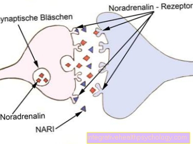
All of this information reaches the cerebellum from the spinal cord, vestibular nuclei and cerebral cortex via so-called moss fibers that end at the granule cell layer. The granule cells are excited by these endings and in turn excite the Purkinje cells (as already mentioned, the granule cells are the only excitatory nerve cells in the cerebellum, they use the neurotransmitter glutamate). Since the Purkinje cells have an inhibitory effect, this would mean that the Purkinje cells simply massively inhibit everything that they can achieve with their cell processes. But that would not be helpful for the functionality of our motion sequences. And so the other inhibitory cell types in the cerebellum now come into play. Star cells, basket cells and golgic cells have an inhibitory effect on the Purkinje cells in various ways (shown in simplified form in the diagram). What results from this is an inhibition of inhibition, which means something like a certain, but not too strong, excitement. To understand what is being excited in this way, one has to look at the top part of the diagram. The cerebellum sends information to the spinal cord, vestibular nuclei and cerebral cortex via the Purkinje cells. To do exactly what was described above. Coordinating head and body posture, coordinating eye movements and directing movements in the exact direction and not choppy, but fine-tuning.
The cerebellum plays a key role in implicit learning. Well-trained movement sequences are “stored” in the cerebellum; you no longer have to think about it while performing them. Think, for example, of cycling or driving, playing the piano and dancing.
Also read our article: motor learning


.jpg)