Cornea of the eye
introduction
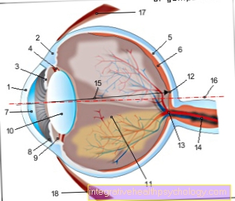
The cornea (Cornea) covers the anterior segment of the eye. This is an approx. 550 micrometer to 700 micrometer thin, transparent, collagen-containing layer that cannot be seen with the naked eye.
It serves to protect the eyeball and refract the incident light rays.
Structure of the cornea
The cornea consists of several layers (structure).
The multilayered corneal epithelium protects the corneal surface and repels germs. Together with the tear fluid, it forms the smooth, refractive surface of the optical system. The basal epithelial cells are anchored in a basement membrane, which is embedded in the so-called Bowman membrane (a thicker and coarse layer) and contributes to the stability of the cornea.
The corneal stroma is formed by parallel layers of collagenous fibers and is transparent due to its regular and narrow lattice structure.
On the inside of the cornea (Cornea) is the single-layer corneal endothelium. Its basement membrane is also traversed by elastic fibers and is called the descement membrane. The corneal endothelium seals the corneal stroma from the aqueous humor. Any liquid that has penetrated is pumped back into the anterior chamber.
The cornea is not able to form again after deeper injuries. The structure of the cornea remains permanently damaged.
You may also be interested in this topic: Anatomy of the eye
Functions of the cornea
First of all, the Cornea as a front lens, i.e. it contributes to the imaging of the image on the retina with its own refractive power. Its refractive power is 43 diopters.
In addition to its contribution to vision, the cornea also has a protective function. In this way, it can cushion the intraocular pressure generated in the eye. The cornea is a very important part of the optical apparatus that cannot be done without.
Also read our articles:
- How does tendons work?
- Visual acuity

- Cornea - Cornea
- Dermis - Sclera
- Iris - iris
- Radiant Bodies - Corpus ciliary
- Choroid - Choroid
- Retina - retina
- Anterior chamber of the eye -
Camera anterior - Chamber angle -
Angulus irodocomealis - Posterior chamber of the eye -
Camera posterior - Eye lens - Lens
- Vitreous - Corpus vitreum
- Yellow spot - Macula lutea
- Blind spot -
Discus nervi optici - Optic nerve (2nd cranial nerve) -
Optic nerve - Main line of sight - Axis opticus
- Axis of the eyeball - Axis bulbi
- Lateral rectus eye muscle -
Lateral rectus muscle - Inner rectus eye muscle -
Medial rectus muscle
You can find an overview of all Dr-Gumpert images at: medical illustrations
Corneal diseases
Astigmatism
A Astigmatism is also called astigmatism designated.
This is a harmless and very widespread corneal anomaly that can be observed in around 70% of all people who wear glasses. Literally translated, astigmatism means “pointlessness”. In German, astigmatism is also called “astigmatism”.
A normal and healthy cornea has a uniform curvature in all directions of its radius. In people with astigmatism, which is usually congenital and does not recede in the course of life, the cornea is now slightly more curved in one direction than in the other. As a result, the light rays hitting the eye are no longer shown as punctiform, but rather as lines on the retina.
The horizontal rays of light are refracted more than the vertical ones. As a result, the rays do not come together in a single sharp focus on the retina. Instead, two different rod-shaped focal lines are created: the image appears slightly distorted. This explains the term “astigmatism”.
Astigmatism can very often be observed in combination with other refractive errors of the eye, for example in combination with nearsightedness or farsightedness.
Once astigmatism is recognized and diagnosed, it can be easily remedied with glasses, contact lenses or refractory surgery on the cornea.
Read more about the topic here:
- Symptoms of astigmatism
- Laser therapy for astigmatism
Inflammation of the cornea
Inflammation of the cornea is also known as keratitis. It can have many different causes, but most of the time it is an infection. Various pathogens such as bacteria or viruses, but also fungi can occur here.
Non-infectious causes can be, for example, strong UV light or insufficient eyelid closure, which leads to the cornea drying out.
Because the cornea of the eye is very sensitive, corneal inflammation can cause severe pain. In addition, there is often a reddening of the eye, which can secrete secretions or tears. This is especially the case if there is inflammation of the conjunctiva at the same time.
Corneal inflammation is a serious disease that should be treated. Otherwise the cornea may become cloudy or scarred.
The most common form of corneal inflammation is bacterial infection. It can occur when wearing dirty contact lenses. Any pathogens adhering to it can attack the eye. A purulent secretion is typical of bacterial inflammation. Bacterial corneal inflammation is considered an emergency as it can go as far as the perforation of the protective cornea.
You may also be interested in the following topics:
- Eye infection
- Injury to the eye from rays
Corneal detachment
The cornea of the eye forms the front wall of the eyeball. Behind her lies the anterior chamber of the eye. This is the space between the cornea and the iris. The cornea does not lie on any tissue from which it could detach. Not to be confused with a detachment of the cornea is a detachment of the retina, which, however, is located in a different place in the eye.
The cornea of the eye consists of several different layers of tissue. On the inside of the cornea there is a thin layer of cells, which is also called Endothelium referred to as. In the Fuchs endothelial dystrophy With increasing age, these endothelial cells die down. This presumably has, among other things, hereditary causes. The endothelial cells are required for the metabolism of the cornea. As a result, the cornea swells. Cell layers can stand out here. The cornea can also become cloudy.
Gaps within the cornea are also conceivable as a result of injuries to the eye.
More on this: Corneal detachment on the eye
What do you do if you injure your cornea?
First aid for an injury to the cornea always depends on the type of injury.
A common cause of corneal injuries are foreign bodies that can arise from improper grinding or drilling. If such foreign objects penetrate the cornea, it can be very difficult to determine the severity of the injury. Therefore, one should consult an ophthalmologist if the cornea is injured. This can safely remove any foreign body that may still be in the eye.
In addition, an antibiotic is often given as an eye drop, for example. This is especially necessary if dirt has been brought into the eye with the foreign body or if there is a defect in the cornea through which pathogens can penetrate.
But not only mechanical force can damage the cornea. Many harsh chemicals can also damage the cornea. The first aid in this case is to thoroughly rinse the eye with clean water. This should be done as soon as possible.
Many workplaces also have special devices for rinsing the eye. In addition, an ophthalmologist should be consulted immediately.
You can find more here on the topic:
- Injuries to the eye
- Burns to the eye
Corneal transplant
Corneal transplantation can be performed if corneal diseases severely limit the eye's vision, or corneal diseases that cannot be controlled otherwise. This involves removing the patient's cornea and replacing it with a donor cornea.
It is possible to replace the entire cornea or individual sections. It is a corpse donation, so the cornea is only removed after the donor's death.
The cornea is usually not supplied with blood. This has several advantages in corneal transplants. Since the cells of the cornea do not depend on blood flow, they remain viable for days after the donor's death and can theoretically be transplanted. In addition, the recipient's immune system usually does not react to the foreign tissue because it does not come into contact with it due to the lack of blood vessels.
Read more about the topic here: Corneal transplant


.jpg)