Scaphoid fracture in the foot
definition
Besides the typical Scaphoid fracture on the wrist a fracture of the foot is also possible.
The Scaphoid bone of the foot is called in the technical language "Os naviculare" and is located between the talus and the sphenoid bones of the first two toes on the side of the big toe.
A Fracture of the scaphoid bone on the foot is rare, but especially less than one Scaphoid fracture of the hand. In case of fractures of the foot, in addition to the toes, mainly these are Ankle bone and the Heel bone affected, fractures of the small ones Tarsus are unusual.

causes
Fractions of the Scaphoid on the foot often arise through direct violence on the area of the bone. It is often a matter of deliberate blows with blunt objects or accidents such as a forceful blow against a ledge.
Falling or unfortunate twisting and defending of the foot can also cause fractures in the smaller bones of the foot.
Since the navicular bone is a very small bone and offers hardly any surface for fractures to attack, the force of force must act specifically on the bone. It is not uncommon for the bones to be through Inflammation of the bones or osteoporosis pre-damaged, which causes instability of the bones.
Symptoms
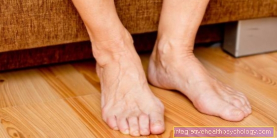
It usually occurs immediately after the fracture local pain of the Inside of the foot. The foot can swell, become warm and hurt when touched and moved.
In addition, bruises can form, which causes the affected area around the navicular bone to turn visible red on the outside.
The swelling and pain are accompanied by a loss of function, making it difficult to step on and walk.
These symptoms can also be a strong contusion act.
Depending on the severity of the scaphoid fracture, the fracture can be feel or there is slight deformation.
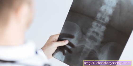
In order to be able to distinguish a bruise from a fracture with certainty, only one helps X-ray image. As a rule, the breaks can be recognized and reliable diagnoses made.
Appointment with ?

I would be happy to advise you!
Who am I?
My name is dr. Nicolas Gumpert. I am a specialist in orthopedics and the founder of .
Various television programs and print media report regularly about my work. On HR television you can see me every 6 weeks live on "Hallo Hessen".
But now enough is indicated ;-)
Athletes (joggers, soccer players, etc.) are particularly often affected by diseases of the foot. In some cases, the cause of the foot discomfort cannot be identified at first.
Therefore, the treatment of the foot (e.g. Achilles tendonitis, heel spurs, etc.) requires a lot of experience.
I focus on a wide variety of foot diseases.
The aim of every treatment is treatment without surgery with a complete recovery of performance.
Which therapy achieves the best results in the long term can only be determined after looking at all of the information (Examination, X-ray, ultrasound, MRI, etc.) be assessed.
You can find me in:
- Lumedis - your orthopedic surgeon
Kaiserstrasse 14
60311 Frankfurt am Main
Directly to the online appointment arrangement
Unfortunately, it is currently only possible to make an appointment with private health insurers. I hope for your understanding!
Further information about myself can be found at Dr. Nicolas Gumpert
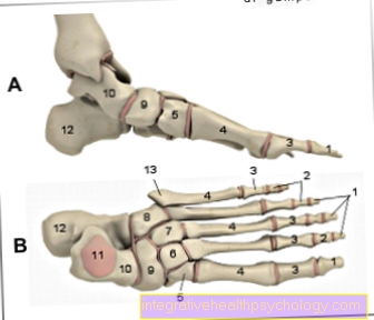
- Toe phalanx -
Phalanx distalis - Middle toe -
Phalanx media - Phalanx -
Phal. proximalis
(1st - 3rd toe bones -
Phalanges) - Metatarsal bones -
Os metatarsi - Inner sphenoid bone -
Medial cuneiform bone - Middle sphenoid bone -
Os cuneiform intermedium - External sphenoid bone -
Os cuneiform laterale - Cuboid bone - Os cuboideum
- Scaphoid bone - Navicular bone
- Ankle bone - Talus
- Ankle roll - Trochlea tali
- Heel bone - Calcaneus
- Protrusion on the 5th metatarsal -
Tuberositas ossis metatarsalis quinti (V)
You can find an overview of all Dr-Gumpert images at: medical illustrations
therapy
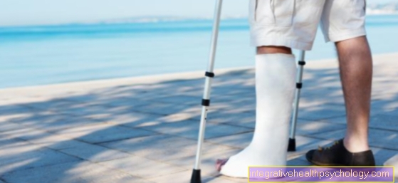
Treatment of a scaphoid fracture is usually given conservative. If the bones have not shifted against each other and do not restrict the movement of the foot, a plaster created. This immobilizes the foot for several weeks in everyday life to prevent the fracture from worsening and to ensure that it heals.
Physiotherapy can be used to encourage movement of the entire foot. However, the load on the navicular bone must be kept as low as possible.
If the break is postponed or the bone shattered, a surgery take place, in which the bone parts are put together in the right place and fixed to one another with wires.
Afterwards, a plaster cast must also be worn to immobilize the fracture so that the break heals in peace. Such operations are nowadays often minimally invasive as part of a Arthroscopy carried out.
Duration
Since the foot is loaded with the full body weight with every step, the bones are exposed to great pressure. It takes a long time for the scaphoid fracture to heal completely Immobilization respectively.
With a normal healing time you have to go with about 6-8 weeks calculate before the foot can be fully loaded again. After an operation, this healing time is often extended by two weeks.
In rare cases, excessive muscle wasting after the immobilization phase requires a protracted one Muscle building kick off. Osteoarthritis changes can also be chronic consequences of a broken bone (see: Pseudoarthrosis)