Hollow of the knee
definition
As the back of the knee or also fossa poplitea is an anatomical structure on the back of the knee. It is diamond-shaped and is outwardly through the Biceps femoris muscle - limits the two-headed thigh muscle. The semimembranosus and semitendinosus muscles join inwards, i.e. towards the middle of the knee. Both ensure flexion and internal rotation of the knee joint. Your tendons can be felt inside the hollow of the knee when the knee is tensed. The two heads of the gastrocnemius muscle, i.e. the calf muscle, limit the hollow of the knee towards the bottom. The muscles together form a diamond in which several important anatomical structures run.

Anatomy of the hollow of the knee
Several run through the hollow of the knee annoy and Vesselssupplying the lower extremity. The sciatic nerve belongs to them, or Ischadic nervewhich supplies most of the larger muscles along its supply route from the tailbone to the heel. The sciatic nerve is considered to be the strongest and thickest nerve in the body. It runs from its origin in the sacral nerve plexus along the back of the thigh, crosses the knee flexors, and then splits into the hollow of the knee at the level of the hollow of the knee Tibial nerve and the Common fibular nerve on. These in turn supply the Lower leg muscles.
In addition to the sciatic nerve, they pierce in the hollow of the knee Vena and Popliteal artery the knee. Both run as first Vena and Femoral artery on the front of the thigh until they migrate through the so-called adductor canal to the back of the knee into the hollow of the knee. From this point onwards, their new anatomical names will also be assigned to them. The popliteal artery soon divides again into anterior and posterior tibial arteries.
Likewise, there are lymph nodes in the hollow of the knee, which are known as Nodi lymphoidei are designated. A distinction is made between deep and superficial popliteal lymph nodes. The hollow of the knee is covered on the outside by a thin layer of skin that is sensitively supplied by several nerves.
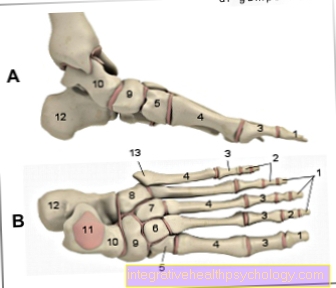
Illustration popliteal pain

Back of the knee pain
A - tear of the outer band / inner band
B - injuries to the menisci
C - osteoarthritis
D - popliteal cyst / Baker's cyst
E - thrombosis
- Inner meniscus -
Medial meniscus - Inner band -
Ligament collateral tibial - Popliteal muscle -
Popliteus muscle - Shin - Tibia
- Internal calf muscle -
M. gastrocnemius, caput mediale - External calf muscle -
M. gastrocnemius, caput laterale - Two-headed hamstrings
Biceps femoris muscle - Half-tendon muscle - Semitendinosus muscle
- Femur - Femur
- Posterior cruciate ligament - Posterior cruciate ligament
- Articular cartilage - Cartilago articularis
- Anterior cruciate ligament - Ligament cruciatum anterius
- Outer meniscus - Lateral meniscus
- Outer band - Ligamentum collaterale fibulare
- Fibula - Fibula
All medical illustrations
Pain in the back of the knee

Pain in the hollow of the knee can have a wide variety of causes, as a number of important anatomical structures run here. Pain can also radiate into the thighs and lower legs, originate from the front of the knee, or be part of a vascular disease.
Is the back of the knee swollen and hurts, this can be done, for example, on a Strain in surrounding muscles lie. A strain is often the result of excessive exercise and will resolve itself after a few days. A typical strain is that it load-dependent is and quickly disappears if you take care of yourself. It is different with one Meniscus damagewhich can also take a chronic course over months and years. Since the menisci in the knee act like a kind of cushion between the thigh and lower leg bones, damage primarily occurs Load and rotational movements noticeable in the knee joint. This sharp pain usually affects the knees, but it can also hurt the hollow of the knee.
At Athletes One should also think of otherwise rather improbable causes: For example, a hypertrophied, so Press the greatly enlarged muscle on the popliteal artery and compress it. Runners and cyclists often experience one Irritation of the muscle-tendon transition in the thigh muscles. Typically, tenderness occurs when palpating the M. biceps femoris–Head up. Stretching the leg also hurts.
Back of the knee taping
For some years now, you have seen more and more athletes walking around with adhesive tapes in the brightest colors. But what is a Tape well, and can it help with knee and hollow knee pain?
First of all, you have to choose between "Kinesio tapes" and "normal“ Tape distinguish which one is also used in the hospital. The latter is often used in martial arts to Stabilize joints. In boxing matches, the wrists are wrapped so that they do not twist and break when punched. In sports such as judo or karate, you can often see taped ankles, because in these sports you twist your ankle particularly often. So one can tap stabilizing and preventive measure be.
It is to be distinguished from the "Kinesio tape", Which is not wrapped around joints in many layers, but is usually stretched over a large area of one or two layers over muscle parts and is available in different colors. According to the manufacturer, the colors should have a psychological effect, for example a blue tape should cool, while a red tape should create a feeling of warmth. Tapes can do that Stabilize the knee joint, but also at Muscle tension, or strains shorten the healing time significantly. They adhere very firmly to the skin and massage the muscle with every movement. This stimulates the flow of blood and lymph, relieves the muscle, and relieves the pain. The hollow of the knee itself is difficult to access for a tape bandage, but the surrounding muscles that form the hollow of the knee can be taped. The mobility of the knee is not restricted, the muscles are even relieved, which can lead to a decrease in muscle mass. At this point, however, this is beneficial as the muscle can be rebuilt after it has healed.
Popliteal cyst
If the back of the knee is swollen, it may be Baker's cyst act. This cyst was originally called the popliteal cyst - due to its anatomical location - but is now named in honor of its discoverer William Baker, an English surgeon in the 19th century. It is a Protuberance of the posterior wall of the Knee joint capsule between the two muscles M. semimembranosus and M. semitendinosus. A Inflammatory response in the knee leads to excessive production of synovial fluid, as a result of which the joint capsule expands. She evades to the point of least resistance, in this case the hollow of the knee. This is then swollen, hurts, and can be pressed in softly. The Baker cyst can also press on the vessels and nerves running in the hollow of the knee and impair their function. In extreme cases, there is one Underperfusion the surrounding structures, or too Signs of paralysis of the lower extremity accompanied by severe pain. If left untreated, a Baker's cyst can ruptureAs a result, the synovial fluid is freely distributed in the lower leg. This causes a strong increase in pressure on muscles and vessels, compression and even death of the leg. One speaks of one Compartment syndrome, an absolute emergency indication that must be surgically treated within hours. Of course, not every Baker cyst has to end in a compartment syndrome, but it definitely requires a doctor's examination, as it is usually based on joint damage with inflammation that should be treated.
thrombosis
A particularly dangerous complication of pain in the back of the knee is thrombotic vascular occlusions of arterial or venous nature. Most of the time it is a thrombus, i.e. a clot of blood that attaches to narrow areas in the venous system. Such a thrombus has no contact with the vessel wall, and is also called red (because platelet-rich) thrombus designated. The basis is arteriosclerosis, i.e. vascular calcification, in combination with little movement, or one Narrowing of the vessel. But also smoking, Insufficient fluid intake, and the birth control pill (see: Thrombosis risk of the pill) can promote thrombosis.
A thrombus also develops in one Vascular injurywhen the opened vascular site is to be closed again. To this end, platelets are always circulating in the blood, and they attach themselves to certain factors that are released in the event of a vascular injury. This physiological thrombus is called a white thrombus, or coagulation thrombus. A thrombosis obstructs the vessel, leads to blood congestion and causes it to swell. In addition to overheating and reddening of the pent-up tissue, this also leads to pain due to the increased pressure.
A acute thrombosis mostly arises after long immobilization of the foot, for example on long flights, or after applying a plaster cast. A thrombus can loosen and be flushed with the blood flow into the lungs, where it then cuts off sections of the lungs from the blood supply. This clinical picture, known as pulmonary embolism, is an acute emergency and requires immediate emergency medical care, otherwise parts of the lungs will die. The Popliteal artery relocated by a thrombus be what is going through Coldness, lack of pulse and numbness on the foot and lower leg expresses. The thrombus is not flushed towards the heart here, but relocates the increasingly thinner vessels that pull towards the foot. You can imagine it like a ball rolling along an increasingly narrow tube: At a certain point it becomes stuck and no more blood can pass through. Here, too, emergency medical care is indicated simply because of the pain.
diagnosis
Pain in the back of the knee can have a wide range of causes. An MRI is usually performed to rule out a Baker cyst. 90% of all meniscus damage can also be detected in the MRI. Unfortunately, this procedure is very expensive (€ 1000-2000 per imaging) and is therefore not always the first choice. An orthopedic or physiotherapeutic examination can also provide information about the problem. Thromboses, however, are usually diagnosed very quickly through a clinical examination with palpation of the legs and, if necessary, an ultrasound examination. Muscle fiber tears and strains usually do not necessarily require a medical examination, unless they worsen or become chronic. Here, gently palpating and massaging the affected area can accelerate the healing process.
prophylaxis
Proper warm up, a adequate body weight, and correct execution of the exercises is the be-all and end-all to prevent problems in the knees and back of the knees. Prophylactic can also stabilizing tapes (e.g. "Leukosilk"), or kinesio tapes can be used. In order to prevent deterioration in the event of an injury, one should adhere to the PECH scheme Hold (pause, ice, compression, elevation).