Bone marrow edema
introduction
As Bone marrow edema syndrome (KMÖS) or Transient osteoporosis This is a temporary disease of the bones, in most cases the hip. knee and upper hocks however, can also be affected, albeit less often. A spontaneous pain in the hip is the classic leading symptom of this disease. Men are statistically affected significantly more often than Women. The disease usually occurs in both sexes middle age, i.e. between the 3rd and 5th decade of life. A diagnosis can be made based on the symptoms and by means of MRI very safe.
causes
The causes of the primary bone marrow edema syndrome have not yet been clarified, which is called "idiopathic" referred to as.
However, it can occur secondarily as a result of other diseases.
Above all, traumatic injuries, such as bruises, play a direct role or can indirectly lead to tissue atrophy via circulatory disorders and ultimately to a BMO.
Even in the last trimester of pregnancy, BMES can occur in rare cases due to compression fractures of the lumbar spine.
diagnosis
X-ray examinations are with primary Bone marrow edema syndrome is usually normal, as a reduction in bone density is only visible after a loss of 40% of the usual bone substance. Only sometimes, but usually only one to two months after the onset of the symptoms, is a focal (focal) To recognize density decrease. The secondary BMES, on the other hand, can show characteristic changes in the underlying disease on X-rays. The inflammation and rheumatism values in blood tests remain negative for both forms.
The best way to diagnose bone marrow edema with almost 100% certainty is to use an MRI scan and to distinguish it from other diseases. This shows clear bone marrow edema, i.e. the increased accumulation of tissue fluid, especially in the femoral head and femoral neck. This can also be the case in deeper regions of the thigh bone and appear as a blurred area. This image is typical of a KMÖS. Scintigraphy can also be useful in making the diagnosis. By using radioactive markers, a characteristically increased blood flow to the hip and increased activity of the bone-forming cells become visible.
The most important differential diagnosis is osteonecrosis. This is the death of bone substance as a result of an infarction (the Closure of a vessel). However, osteonecrosis can be clearly distinguished from BMES with the above-mentioned examination results.
Symptoms
The clinical picture of the Bone marrow edema syndrome is characterized by acute stress pain in the groin and a limping gait pattern as a result of this. The intensity of the pain usually increases over time, but in any case it will not go away completely. Pain at rest and at night usually do not occur. Also typical are Restrictions on movement in the area of the hip joints. Above all, splaying, bending the hips and rotating the thigh around its own axis are difficult.
therapy
The aim of treating bone marrow edema is to achieve partial or total discharge the hip, as well as Freedom from pain. This is through the administration of medication like Ibuprofen or Diclofenac (to the group of NSAIDs due) and sometimes with weak opioids, such as tramadol. With the help of physical therapy can also Micro- and Compression fractures of the only slightly resilient bone can be prevented. Furthermore, the intake of Aminobisphosphates promote bone formation. These have essentially two effects on the bone. On the one hand, they are made up of cells that break down bone substance, the Osteoclasts, absorbed and inhibit their activity, on the other hand they can attach to the surface of bones and thus directly to Mineralization contribute. In contrast, the gift of Calcitonin (a endogenous hormone to build up the bone) and Cortisone not proven. Prostacyclin and structurally similar active ingredients can partly be used in off-label use, i.e. without clinically proven proof of effectiveness, and achieve a positive effect. A Drilling of the edema, as it is often done in osteonecrosis, can be done by a Pressure relief of the bone and the subsequent improvement in blood circulation lead to an immediate and significant improvement in symptoms. In each case, however, is one medicinal and physiotherapeutic Therapy of the highest priority.
prophylaxis
As with other musculoskeletal disorders Sports is an extremely good means of preventing bone marrow edema. By building up the muscles, a good stability of the joints and thus the risk of injuries to the bones and the chance of falls is significantly reduced. Also promotes the stress on the bone metabolism and thus the construction of the Bone substance.
According to location
Bone marrow edema of the knee
A person's knee does not consist of a single bone, but should be viewed as a joint. It consists of three bones that are stabilized with the help of ligaments and muscles. Its bony parts include the thigh, the shin and the kneecap.
If fluid accumulates in one or more bones of the knee joint, it is called bone marrow edema of the knee.
- Root cause:
The cause of the accumulation of fluid in the small cavities of the bone can range from an accident to a metabolic disease and must be carefully clarified by a doctor. - Symptoms:
Those affected feel pain in the knee area symptomatically, which can be explained by the increased pressure on the bone structures.
The liquid fills the small spaces between the bone and exerts an unusual force on it from within. The pressure increases when the affected joint is stressed, when your own body weight and gravity put additional stress on the joint.
Movement-dependent pain peaks are therefore typical of bone marrow edema in the knee with an otherwise normal appearance of the joint.
The fluid in the bone is invisible to the naked eye from the outside. From a purely visual point of view, it can therefore correspond to a healthy knee. - Therapy:
The therapy always depends on the cause of the bone marrow edema, but usually includes an initial protection of the affected joint. Relief with forearm crutches is sufficient and maintains the mobility of the joint. With the protection you enable the body to get the inflammatory reaction under control.
Movement would only mean further irritation of all structures and reactively lead to even more fluid accumulation.
Lymphatic drainage can help drain the fluid even faster.
How long the relief of the knee joint should take depends on the healing process. Experience has shown, however, that it should last about six weeks and be replaced over the course by gradually reloading.
Bone marrow edema takes a long time because the metabolism of the bones slows down with age.
Repair processes therefore require more and more time with increasing age. It often takes about a year for bone marrow edema to heal completely.
For athletes, this means gradually increasing performance and not doing competitive sports for at least three months.
If those affected put pressure on the joint too quickly, the bone threatens to lose stability and elasticity.
The more pressure is exerted on the bone by the accumulated fluid, the more its small blood vessels and nerves are compressed. The result is an undersupply of the bone tissue, which in the worst case shows up in the loss of the bone. Broken bones could result.
In order to avoid excessive stress, painkillers should be used in sufficient amounts but in doses. Only in extreme cases should operational measures such as a relief well be considered. They often involve unnecessary risks and do not shorten the course of the disease.
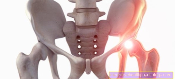
Bone marrow edema of the hip
Bone marrow edema of the hip is an accumulation of fluid in the small cavities of the hip bones.
The liquid is preferably stored in the femoral head.
- Symptoms: To better understand the symptoms, it is important to know the structure of the hip joint. It consists of the thigh and the pelvis. So that these two bones form a joint, the thigh has a femoral head and the pelvis has a joint socket. Only with a perfect fit do they result in an optimal joint together with ligaments and muscles.
If fluid accumulates in the femoral head, the fluid presses on the plexus of its bone structures. The result is a painful inflammatory reaction in the bone itself. If the leg or hip is not moved, it does not hurt because there is no friction point between the hip bones in a calm position.
However, if the person puts pressure on the leg, this leads to pain. The joint surfaces meet and the nerves in the femoral head are compressed and irritated by the pressure of the fluid and the body's own weight.
The pain radiation often extends into the groin and often leads to a limp. - Causes: The cause of bone marrow edema in the hip must always be found out on a case-by-case basis.
Often, age-related osteoarthritis in the hip is the trigger for fluid build-up. Read more about the topic here: Hip arthrosis
In the same way, even the smallest bone fractures or metabolic diseases such as rheumatoid arthritis can trigger bone marrow edema. - Pregnancy: If it occurs temporarily during pregnancy, doctors speak of pregnancy-associated osteoporosis. It occurs more frequently in the last third of pregnancy in first-time mothers. The cause has not yet been clarified.
However, a connection with the changed hormonal influences during pregnancy is suspected. - Therapy: Immobilization is the first choice for any bone marrow edema.
Those affected must therefore relieve their affected leg with crutches and reclining phases for at least three to six weeks.
In order to keep the hip joint flexible, physiotherapy should be carried out after the acute phase. As it heals, it should increase in intensity and duration. Since bone marrow edema takes a long time to heal, physiotherapy can take months.
Pain relievers may be taken to relieve the pain.
Further therapy depends on the cause of the disease and aims to eliminate the trigger. Complete freedom from symptoms can usually be expected after a year, but in individual cases it can be longer.
Bone marrow edema on the shoulder
Bone marrow edema of the shoulder is often the result of accidents or age-related wear and tear on the bones.
- Causes:
Both causes cause irritation of the bone and reactively lead to an inflammation-related accumulation of fluid in its interstices and especially in its bone marrow. The fluid helps the body heal the inflammation better.
The blood vessels become more permeable in the course of wound healing at the inflamed area and allow cells and valuable substances to migrate into the tissue so that they can take over their defense and repair functions.
The fluid in the form of bone marrow edema is therefore accumulated by the body itself. The bone is a very strong tissue that cannot stretch as well as the skin.If too much fluid accumulates in its cavities, it presses on its solid structures as well as on its blood vessels and nerves. The effect is the sensation of pain. The pain will only subside when the fluid in the bone is reduced.
- Therapy:
Protecting the shoulder is therefore essential for therapy. The relief minimizes the inflammatory stimulus and enables the fluid to be transported away again via the lymph and blood vessels.
If the person concerned were to put additional strain on their shoulder, the pressure on the bones would additionally irritate the nerves and the body would try to bring even more fluid with defense cells into the inflamed area.
Carrying and lifting loads and exercising must therefore be avoided for at least three weeks (in some cases up to six weeks).
Depending on the sensitivity to pain, this is followed by partial exertion, which must be increased in steps up to full exertion.
With the shoulder, it is very difficult to estimate when complete healing can be achieved.
In order to maintain the mobility of the joint in all degrees of freedom, it must be moved regularly. The movement, of course, always irritates the joint and delays the healing process. Nevertheless, it is necessary in order not to retain any restrictions later.
Physiotherapy is therefore best recommended for adequate stress. It can take up to a year for complete healing.
Bone marrow edema of the ankle
Ankle bone marrow edema is an accumulation of fluid in one or more bones of the ankle.
- Causes:
Often the cause is trauma, which not infrequently happens during exercise.
The irritation of bone structures can occur suddenly as a result of incorrect loading or permanently due to overload. So if someone twisted their ankle they might as well develop bone marrow edema as if they overexert their bones from running for days.
Metabolic diseases, medication or impaired blood circulation in the ankle area can also cause bone marrow edema in the ankle. - Symptoms:
The symptoms are always comparable and are mainly shown by pain in the ankle. The ankle may initially be swollen from the trauma, although swelling is not a characteristic sign of bone marrow edema.
It is much more typical that those affected feel pain without a cause being recognizable from the outside. The pain occurs especially during exercise and increases in intensity.
Depending on the location of the bone marrow edema, the pain can also extend into the foot or shin. - Therapy:
Usually immobilizing the ankle is enough to heal the bone marrow edema. Those affected are often initially given forearm crutches for complete relief and then a splint or roll-off aid for partial load.
The downside is that this treatment method can take up to a year. However, it is also the safest and the least complicated because it is not associated with any surgical intervention.
Surgery is only necessary in rare cases with bone marrow edema. If the pressure from the liquid on the bone is too strong and the bone threatens to break, a relief hole can be made. The fluid can drain through the drilled hole and the pain subsides.
Nevertheless, after such an operation, the ankle must be relieved and the course of the disease is not shortened.
It only protects against bone destruction in an emergency. The procedure itself does not take long if no other structures such as muscles or ligaments have to be corrected by trauma in the same procedure.
Bone marrow edema of the lumbar spine
The diagnosis of bone marrow edema of the lumbar spine is an accumulation of fluid in the bone marrow of one or more vertebral bodies.
There is usually no free fluid in the small spaces between the bones, so edema is always abnormal.
- Causes:
The cause can be varied. Often it is trauma that leads to a contusion of the spine. Metabolic diseases such as those of the rheumatic type or age-related signs of wear and tear can also lead to bone marrow edema of the lumbar spine. - Diagnosis:
It is important to find out how many vertebral bodies are affected by edema and whether the spine is stable.
It is not uncommon for bone marrow edema to occur as a result of displacements or misalignments of the vertebral bodies.
Imaging in the form of magnetic resonance imaging of the lumbar spine provides information about the extent and severity.
The treating doctor then uses imaging to classify the bone marrow changes into different types.
The current Modic classification distinguishes three types.
Type I stands for bone marrow edema. Sometimes it is also given as a modic character. In type II the blood-forming bone marrow is replaced by fat marrow and type III stands for a hardened bone marrow. - Therapy:
Treatment also depends on the cause and change in the bone marrow edema. Basically, however, there is always adequate pain therapy, which should be accompanied by protection of the spine. As a result, lifting and carrying heavy objects and exercising should be avoided.
Depending on the severity, surgery may even be necessary to restore the stability of the spine. If, in addition to back pain or loss of sensitivity, this indicates an entrapment of nerves, which must be resolved as quickly as possible by an intervention.
Bone marrow edema of the cervical spine
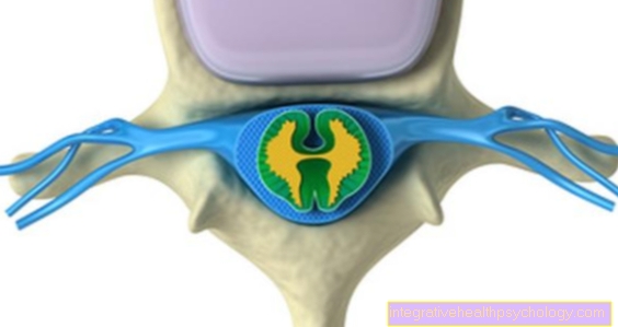
In the case of bone marrow edema of the cervical spine, the solid bone structure of the vertebral bodies contains fluid that can exert pressure on the surrounding structures.
- Symptoms:
If nerves or blood vessels are pinched off, this leads to the development of characteristic symptoms. Those affected feel bone marrow edema of the cervical spine mainly through pain in the neck area, which can even radiate into the shoulder.
But headaches can also occur if the muscles harden as a result.
If loss of sensitivity such as tingling occurs, this indicates an entrapment of nerves.
- causes
There are many causes that can lead to bone marrow edema. The trigger must always be found individually.
Quite often, however, it is trauma or rheumatic diseases that lead to bone marrow edema.
- Diagnosis
To confirm the suspected diagnosis, a doctor will request an imaging of the spine. The images of the magnetic resonance tomography of the cervical spine then make it possible to objectify the severity and the number of affected vertebrae.
In this context, the Modic findings are also collected.
The Modic classification divides bone marrow changes in the area of the vertebral bodies into three types.
Type I stands for bone marrow edema and can also be referred to as Modic's sign.
Type II has a fat marrow instead of the blood-forming bone marrow.
And in type III the bone marrow is hardened.
- Therapy:
Therapy will also be given depending on the cause and extent of the bone marrow edema. If the spine is stable, conservative treatment is possible.
So pain is relieved with medication and the spine is relieved as much as possible. For those affected, this means carrying or lifting as little as possible and not doing any sport. If the pain subsides, the load can slowly be increased again.
However, it often takes about a year for bone marrow edema to heal completely. However, after six weeks of consistent rest, an improvement should already be felt.
forecast
Despite extensive drug therapy and physiotherapy, patience needed when it comes to healing Bone marrow edema goes. Symptoms persist for at least 4 weeks and often up to 6 months. Even if a longer course of the illness of 12 or 18 months is also possible, a chronification of the symptoms is still not known. Whether and to what extent the bone marrow edema syndrome can be viewed as a preliminary stage of osteonecrosis is controversial.