cartilage
Synonyms in a broader sense
- Cartilage cell
- Chondrocyte
- arthrosis
English: Cartilage
definition
cartilage is a special form of connective tissue. A distinction is made between different forms of cartilage, which are adapted to the respective function.
The most important task is the cartilage as a joint surface in the joint and the Intervertebral disc.
introduction
Cartilage is mainly found in the skeleton and in the airways.
Due to its structure and its physical and chemical properties, it occupies a middle position between connective and bone tissue. It has a high compressive strength, is viscoelastically deformable and has a high resistance to shear forces.
Cartilage cells (chondroblasts and chondrocytes) are characteristic of cartilage tissue. These are more or less rounded and lie in small groups (chondrons) directly in the cartilage (in a so-called extracellular matrix) so that they have no contact with one another.
The cartilage cells are equipped with the usual cell organelles. What is noticeable here are the many glycogen particles for anaerobic energy production, i.e. energy production without oxygen) and occasionally individual large fat droplets. This is important because the cartilage is usually not supplied with blood and therefore has little oxygen available.
The most important components of the actual cartilage substance in which the cartilage cells are located - the extracellular matrix - are proteoglycans and collagen fibrils. Both substances are specialty substances that only occur in this form in cartilage.
The compressive elasticity of the cartilage tissue comes from the interaction of the proteoglycans and the collagen fibers.
In adults, the cartilage is free of blood vessels. The necessary nutrients are supplied exclusively via diffusion either via a vascular cartilage skin (perichondrium) or directly via the joint fluid (synovia).
Cartilage growth
The Emergence one cartilaginous structure starts with that Connective tissue cells (Mesenchymal cells) close together and close Cartilage cells (Chondroblasts) differentiate.
You then produce Cartilage matrix and thereby become Chondrocytes.
The cells are pushed apart by the increasing cartilage matrix and form collagen fibrils. This process is known as interstitial growth. This leads to rapid enlargement of the cartilaginous structure and takes place mainly in the early phase of cartilage formation and in the growth plate.
After this Completion of interstitial growth the chondrocytes that emerged from the last cell divisions remain lying together in groups. They are only separated from each other by thin matrix membranes.
Chondrocytes of the cartilage tissue no longer divide. On the outside of the cartilaginous structure, mesenchymal cells develop into connective tissue cells (fibroblasts) and form a connective tissue capsule (perichondrium).
Remain on the inner layer of this capsule undifferentiated cells, from which Chondroblasts can emerge and through Accumulation of new cartilage ensures growth.
The attachment from the outside is called appositional growth designated.
Appointment with ?

I would be happy to advise you!
Who am I?
My name is dr. Nicolas Gumpert. I am a specialist in orthopedics and the founder of .
Various television programs and print media report regularly about my work. On HR television you can see me every 6 weeks live on "Hallo Hessen".
But now enough is indicated ;-)
In order to be able to treat successfully in orthopedics, a thorough examination, diagnosis and a medical history are required.
In our very economic world in particular, there is too little time to thoroughly grasp the complex diseases of orthopedics and thus initiate targeted treatment.
I don't want to join the ranks of "quick knife pullers".
The aim of any treatment is treatment without surgery.
Which therapy achieves the best results in the long term can only be determined after looking at all of the information (Examination, X-ray, ultrasound, MRI, etc.) be assessed.
You will find me:
- Lumedis - orthopedic surgeons
Kaiserstrasse 14
60311 Frankfurt am Main
You can make an appointment here.
Unfortunately, it is currently only possible to make an appointment with private health insurers. I hope for your understanding!
For more information about myself, see Lumedis - Orthopedists.
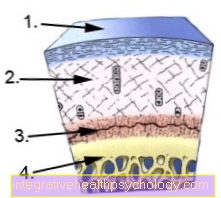
Cartilage build-up
-
superficial layer of cartilage
-
middle layer of cartilage
- calcifying layer of cartilage
- bone
Illustration of a cartilage damage

- Articular cartilage
(hyaline cartilage) -
Cartilago articularis - Cartilage remodeling zone
in bones -
Zona ossificationis - Joint body (articular gnar
of the femur) -
Femoral condyle - Femur -
Femur - Articular cartilage -
Cartilago articularis - Outer band -
Ligamentum collaterale fibulare - Outer meniscus -
Lateral meniscus - Inner meniscus -
Meniscus medialis - Fibula - Fibula
- Shin - Tibia
You can find an overview of all Dr-Gumpert images at: medical illustrations
Forms of cartilage
There are three different types of cartilage tissue:
- the hyaline cartilage
- the fiber cartilage
and - the elastic cartilage
Hyaline cartilage
During the embryonic phase, most of the later bony skeleton in humans is preformed as cartilaginous.
In adolescents, the epiphyseal plates (growth plates) exist within the Long bones out hyaline cartilagewhich is only replaced by bone after growth is complete.
Coated in adults hyaline cartilage the Articular surfaces, forms the sternal part of the Ribs - so the ribs on Sternum -, part of the Nasal septum, the Larynx skeleton and the clasps of the windpipe as well as the big one Bronchi.
You can find everything about hyaline cartilage at: hyaline cartilage
Fiber cartilage
Fiber cartilage occurs in the human body wherever shock absorbers are needed.
Mainly as to:
- of the Spine
- and on Knee joint in the form of meniscus (knee)
- and Discus (Intervertebral disc), the intervertebral discs of the spine,
- in the Symphysis
- the Articular cartilage of the jaw and collarbone sternum joint
- and in the areas of tendons and ligaments that are subjected to pressure.
A decisive difference to the hyaline articular cartilage consists in the amount of collagen fibers. These are significantly increased compared to the basic substance in the fibrous cartilage.
As a result, the fibers are not masked by the basic substance you are already using eye visible. Fibrous cartilage is very similar to taut connective tissue. Because of this, it is sometimes referred to as connective tissue cartilage. The chondrons usually consist of only one or two cells and are arranged parallel to the individual fiber bundles.
Because fiber cartilage both Type I and Type II collagen it contains the transitions to the hyaline cartilage on the one hand and to the tight connective tissue on the other. Adaptation to tensile stress is the focus of fiber cartilage.
Elastic cartilage
Elastic cartilage only occurs in humans:
- in the Epiglottis (Epiglottis)
- in the small Larynx cartilage
- in the auricle
- in part of the external auditory canal
and - in the small bronchi in front.
In addition to the structures in hyaline cartilage there are also elastic fiber nets.
These fiber networks are network-like around the Chondrons created and radiate into the adjacent Cartilage skin (Perichondrium) a. Due to these elastic fibers, it has a yellowish appearance and is significantly more flexible and elastic than the hyaline cartilage. In elastic cartilage there is no mineralization and no bone formation.


.jpg)